Q.6. Describe briefly the structure and life cycle of Chara.
Ans.6. Chara a submerged aquatic algae of fresh water ponds, puddles, tanks, lakes or slow flowing streams. It generally remains attached to the muddy or sandy bottoms with the help of rhizoids. C. baltica occurs in brakish water, whereas C. fragilis occur in hot springs. According to Groves and Bullock Webster (1920) C. fragilis occur in such hot springs where temperature “was such as to boil an egg in four minutes.” Many species of Chara grow in waters running over limestone rocks. Some species become encrusted with calcium carbonate.
Out of about 30 species reported from India, 7, species (Chara grovesil, C. handae, C. hatei, C. nuda, C. psshanii, C. wallichii and C. vandalurensis) are endemic. The most common cosmpolitan species are C. braunii, C. gymnopitys and C. zeylanica.
According to Wood and Imahori (1959), Chara is represented by 188 species in the world.
Structure
The plant body consists of an erect branched axis which may grow to the height of about 20-30 cm. The axis have distinct nodes and internodes. From each node arise a whorl of laterals (branches) of limited growth the so called leaves. From the axis of some of the leaves’, branches of unlimited growth may arise (Thus, the plant body becomes more or less like Equisetum). Each branch, whether limited or unlimited growth, is also differentiated into nodes and internodes. The plant is anchored to substratum by means of colourless, branched, multicellular rhizoids, which arise from the lower nodes of axis.
The growth of axis in length takes place by means of a single dome-shaped apical cell. (BSc Botany Chlorophyta Sample Practice Question Answers)
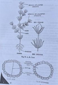
Each node consists of a plate of cells, while the internode consists of single elongated cell which is unsheathed by a cortex of elongated cell. Each cell has a cell wall made up of cellulose and a deposit of CaCO3. They contain single nucleus dense cytoplasm with many discold chloroplasts.
Reproduction
Reproduction is Chara may take place by the following two methods:
(1) Vegetative (2) Sexual
(a) By Amylum stars or starch stars formation: In this method some of the cells of the lower nodes form a mass of special type of cells which are star-shaped and are called amylum starch as they contain amylum starch in their cells. They can give rise to a new plant but their exact mode of development, etc. is unknown.

(b) By bulbils: In this method some of the rhizoids or lower nodes may form bulbils which also give rise to new plants when detached.
(c) By protonema formation: In this method sometimes on the nodes protonema like branches are developed and they also form new plants.
- Sexual reproduction: This is highly advanced oogamous type and takes places with the help of male and female reproductive organs known as globule (antheridium) and nucule (oogonium) respectively. Mostly the species are homothallic or monoecious, a few may be heterothallic or dioecious. The two structures are found just apposed at a node, nucule above the globule.
Structure and development of sex organs
As we know the plant body of chara consists of a main axis which is differentiable into nodes and internodes. From the nodes come out branches of limited growth. These also possess nodes and internodes. The sex organs are developed on the nodes of these branches.
GLOBULE OR ANTHERIDIUM
Structure of the globule
The mature globule or antheridium is circular in outline and red or orange colour. The wall of the globule consists of eight shield cell, the outer wall of each shield ceil is marked with numerous infoldings giving the idea that wall of antheridium consists of more than eight cells but this is only apparent. From the centre of each shield cell there arises a rod like outgrowth, the manubrium which bears at its upper end a primary capitulum cells.
The primary capitulum may from secondary and tertiary cells. The secondary capitulum cells usually bear the branched, uniseriate spermatigenous filaments which are divided into small segments by transverse septa. Each segment functions as a single antheridium. The cytoplasmic contents of each segment give rise to a single spermatozoid or antherozoid. Each antherozoid is spirally coiled and biflagellate.
Development of the globule
Globule, the male reproductive organ, arises in the axis of the branches of limited growth, from the single superficial cell. This call cuts off one or two discoid cell at its basal and then becomes spherical. The lower two cells forms a pedicle while the upper cell enlarges in size and becomes hemispherical in shape. The upper spherical cell divides by two longitudinal and one transverse division to form octant (8 celled structure). This octant divides by two successive curved plates or shields and constitutes the wall of globule.
As the shield cells mature, they develop red pigments and are radial in growths. The mature, shield cells expand laterally and thus a cavity is formed inside the globule. The wall at the base of the globule, is completed by the upper discoid basal cell (pedicle cell) which elongates and protrudes up into the cavity of the globule. The middle eight cells elongates radially to form a rod-shaped manubrium which projects inward from the centre of curved shield cells.
Each of the inner eight cells becomes a primary capitulum. The capitulum cell lies quite close together in the middle of the cavity of the globule.
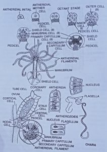
Each primary capitulum cell buds off about 4 to 6 secondary capitulum cells which may further give rise to tertiary and quaternary ones. Now from each capitulum cell there develop antheridial filament. Each antheridial lament consists of upto 200 discoid cells, the antheridium. Within each antheridium is produced a single elongated, spirally coiled and biflagellate antherozoid.
When the globule or antheridium is matured, the shield cells fall apart and the antherozoids are liberated by the gelatinsation of antheridial walls, or through a pore formed in each antheridial cell.
BSc 1st Year Botany Chlorophyta Sample Practice Question Answers
NUCULE OR OOGONIUM
Structure of nucule
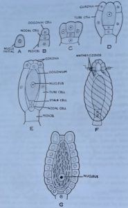
The nucule or oogonium is a short stalked body attached to the body of a primary lateral or leaf or dwarf shoot or branch of limited growth just above the antheridium. A mature oogonium consists of a large oval or elliptical egg surrounded by a cover of five tubular cells which make two or more clockwise spiral turns around it from the upper end of each of the tubular cells, a cell is cut off, thus constituting the crown or corona of oogonium.
Development of nucule
The oogonium or nucule, the female reproductive organ, develops in the axil of the branches of limited growth on the adaxial side. It develops from single superficial adaxial cell. This cell undergoes two successive divisions thus forming three cells. The lower most cell elongates and forms pedicel, the middle cell give rise to five peripheral cells and upper most act as oogonial mother cell.
Each of the peripheral cell divides transversely forming an upper smaller cell corona cell and the lower larger cell tube cell The five corona cells elongates little and mature into the corona. The five tube cell elongated vertically and divides transversely to form a short stalk cell and an elongated oval shaped oogonium containing single uninucleated egg.
The nucule when mature, the tube cells separate, from one another just below corona to form five small slits for the entrance of the antherozoids.
Fertilization
At the maturity of oogonium, the tube cells separate, thus forming slits. The apex of the oogonium gelatinzes. Several antherozoids enter through these slits but, however, only one succeeds resulting in the formation of the zygote.
Germination of Zygote
The zygote, just after formation, secretes a coloured wall around it and undergoes a period of rest within the oogonium. It becomes red in colour, finally changing into black. It falls from the plant and sinks to the bottom of the pond or stream where it germinates after few weeks or more.
At the time of germination, the zygote nucleus migrates to the apical pole and divides thrice into four daughter nuclei, each is haploid in nature. The upper most one nucleus get separated from the rest of three nuclei by the formation of transverse wall. The lower three nuclei degenerate. The on nucleus thus left divides into by a longitudinal division forming protonemal and rhizoidal intitial. They form the protonema and rhizoid respectively. The protonema increases and develops into a young Chara plant.