BSc 1st Year Lower Non-chordates Aurelia Sample Model Practice Question Answer Papers
BSc 1st Year Lower Non-chordates Aurelia Sample Model Practice Question Answer Papers: BSc is a three-year program in most universities. Some of the universities also offer BSc Honours. After getting enrolled for BSc, there are certain things you require the most to get better grades/marks in BSc. Out of those, there are BSc Study Material, BSc Sample Model Practice Mock Question Answer Papers along with BSc Previous Year Papers. At gurujistudy.com you can easily get all these study materials and notes for free. Here in this post, we are happy to provide you with BSc 1st Year Lower Non-chordates Aurelia Sample Model Practice Question Answer Papers.

Index for BSc 1st Year Lower Non-chordates Aurelia Sample Model Practice Question Answer Papers
Structure of Aurelia: Page 1
The receptor organs in Aurelia: Page 2
The life history of Aurelia: Page 3
Q.1. Give an account of the structure of Aurelia and compare it with that of Obelia medusa.
Or
Give an account of the gastrovascular system of Aurelia and the process of digestion.
Ans.1. Habitat
Aurelia is found in coastal waters and has a cosmopolitan distribution. It represents the dominant medusoid stage. The reduced polypoid phase is represented by a trumpet-shaped scyphistoma.
External Features
Shape and size: Aurelia resembles a hydrozoan medusa. The body is à gelatinous saucer-shaped umbrella with tetramerous symmetry. The size usually varies from 4-12 inches.
Coloration: The umbrella is transparent and bluish-white with reddish or pinkish gonads peeping through it.
Structure: The margin of the umbrella is scalloped. It is broken into eight equal lobes by eight notches. Four of them are perradial and the other soul inter-radial. Each notch carries a sense organ, the tentaculocyst or rhopalium, protected by two leaf-like processes the marginal lappets.
The edges of the umbrella between the adjacent notches have a row of closely set hollow tentacles. These are borne by a delicate fold of subumbrella. This marginal flap is known as velarium or pseudovelum. A true velum is absent in Aurelia. The tentacles bear batteries of nematocysts.
Its upper convex surface is the exumbrella surface and its inner concave subumbrella surface. The following structures are found on the subumbrella surface:
1. Manubrium: Hanging downward from the center of the subumbrella surface is an inconspicuous manubrium bearing a four-sided mouth.
2. Oral arms: The four corners of the mouth are drawn into four long delicate oral arms. Each arm bears a ciliated groove that extends midventrally along its length and leads into the mouth. Groove margins are convoluted and studded with numerous nematocysts.
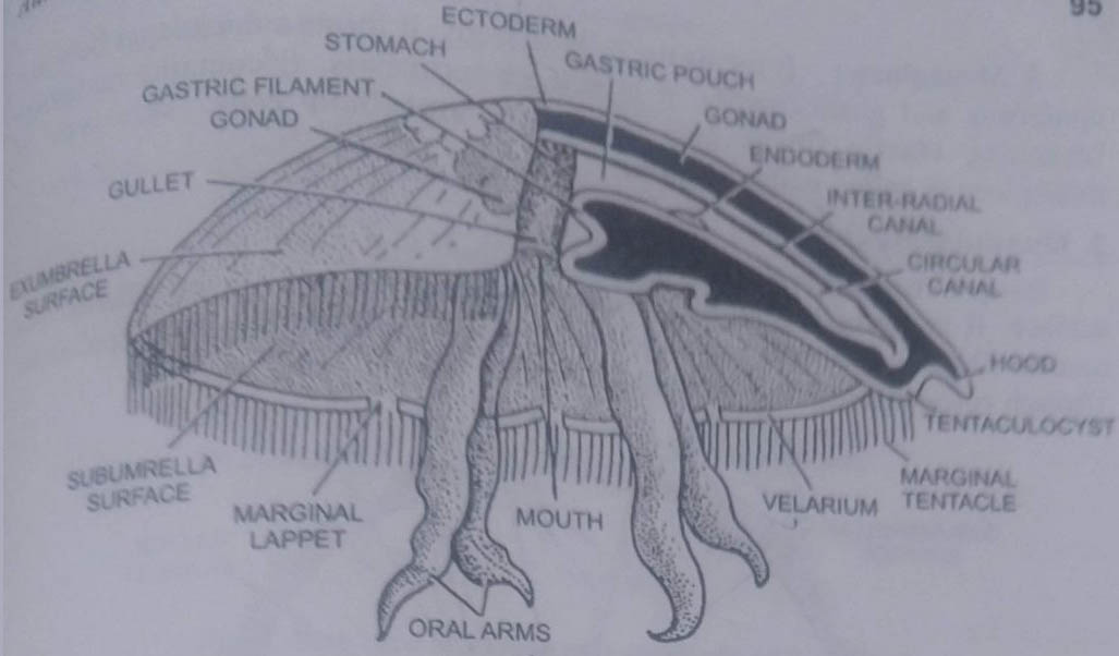
3 Subgenital pits: Situated ventrally between the oral arms and at a short distance from the mouth are four-rounded apertures which lead into four subgenital pits. Each is a shallow cavity just underneath a gonad.
4. The gonads are four horse-shoe-shaped patches of pink or red colour occupying the inter-radial position.
Internal Anatomy
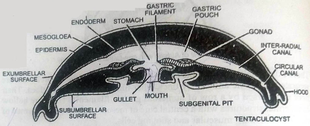
1. Body Wall
The body wall is diploblastic, having the ectoderm and endoderm with a main mass of mesogloea. Their arrangement is similar to the hydroid medusa of Obelia.
1. Ectoderm or epidermis: The exumbrella surface of the umbrella, the velarium, tentacles, the subumbrella surface, the subgenital pits, the oral arms, and the manubrium are all covered with the epidermis. It consists of a single layer of epithelial cells on the exumbrella surface and epithelio-muscular cells on the subumbrella surface. The sensory cells occur between the epithelial cells.
2. Gastrodermis or endoderm: The gastrovascular canals are lined with the gastrodermis. The gastric filaments are formed of a double layer of gastrodermis with a central core of mesogloea. Gonads are also gastrodermal. The cavities of tentaculocysts are also lined by it.
The gastrodermis mainly consist of flagellated columnar endothelial cells. s are also present, but nerve cells and muscle cells are wanting. The endoblasts are restricted to gastric filaments.
3. Mesogloea: It forms the bulk of the body. It forms a thick layer between the epidermis and gastrodermis. It is not structureless. It contains numerous branching elastic fibers and wandering amoeboid cells. This type of mesogloea is called collenchyma.
2. Muscular System
Body musculature is very well-developed and confined to the subumbrella surface. It is formed of both striated and non-striated muscle fibers. The muscle fibers are muscle processes of epitheliomuscular cells of the epidermis. Though ectodermal in origin, these lie in the mesogloea.
The muscle fibres are arranged longitudinally in the tentacles, manubrium, and oral arms but are radial and circular in the umbrella. In the subumbrella mesogloea, the muscle fibres are striated and form a strong, broad, and circular band which is known as coronal muscles. The rhythmic contractions of these muscles help in the swimming movements of the bell.
3. Gastrovascular System and Water Circulation
The digestive system has also taken over the work of the distribution of digested food. Hence a distinct circulatory system is absent and this system is called the gastrovascular system.
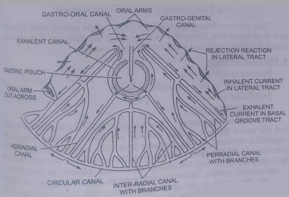
It consists of a rectangular mouth borne at the tip of the manubrium. It leads into the spacious stomach through a short gullet contained in the manubrium. The stomach occupies the whole middle region of the umbrella. It is produced into four wide gastric pouches which are inter-radial in position.
These extend halfway from the centre to the periphery of the umbrella and are separated from one another by thick pillar-like portions of mesogloea. Their floor is traversed by a row of numerous gastric filaments. These are hollow endodermal structures with a central core of mesogloea. These are formed of ciliated, glandular, muscular, and stinging cells.
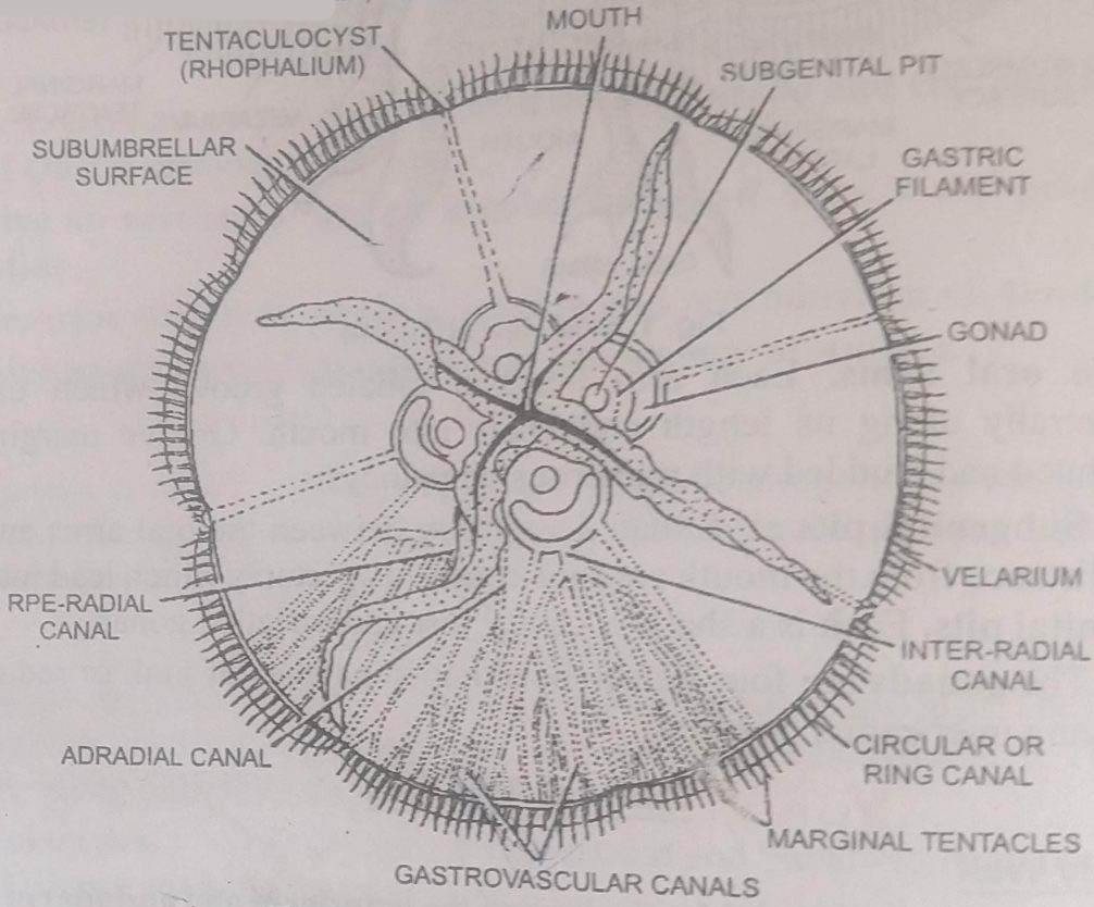
Radial canals: Three radial canals arise from each gastric pouch.
These are :
1. An inter-radial canal in the middle, that immediately divides into three branches. Of them, the outer two produce sub-branches. Finally, all of them join the ring canal.
2. Two adradial canals, one on either side of the inter-radial canal. These do not branch and meet the ring canal.
3. A per-radial canal comes out of an aperture between two adjacent gastric pouches. It branches and finally opens into the ring canal.
Thus there are four-branched inter-radial, eight unbranched adradial, and four branched per-radial canals joining the ring canal or circular canal. The ring canal runs along the margin of the umbrella.
4. Nervous System
The nervous system consists of:
1. Subumbrellar plexus or main nerve net
2. Diffused nerve net
3. Rhopalial ganglia
1. Subumbrellar plexus: It is formed of elongated bipolar nerve cells and the nerve fibers extending over the subumbrella surface between the superficial ectoderm and the muscular layer. This is formed of two distinct plexuses: (i) the sub-epidermal plexus in the manubrium. oral arm, tentacles, and rhopalia, and (ii) the sub-gastrodermal plexus in the wall of the gastrovascular system. The nervous net presents radial thickenings which are strongly developed in the pre-radii and inter-radii corresponding to the position of marginal notches and sense organs. Each is connected with the rhopalial ganglion.
2. Rhopalial ganglia: Special groups of nerve cells are present at the base of marginal sense organs (rhopalia).
3. Diffused nerve net: It lies in the epidermis of the subumbrella as well as the exumbrella. Its nerve cells are small and are connected with the rhopalial ganglia. The diffused nerve net controls local responses like feeding. It also inhibits contractions of the umbrella.
5. Sense Organs
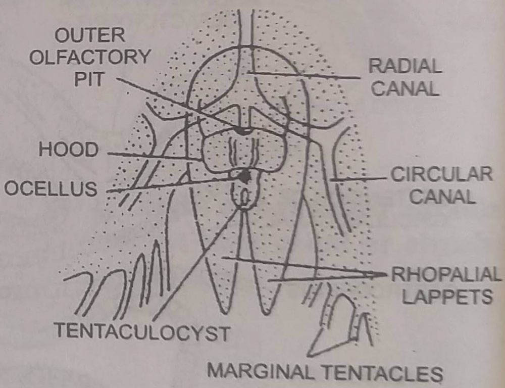
Sense organs of Aurelia are 8 rhopalial situated in the 8 marginal notches. Each rhopalium consists of:
(a) Tentaculocysts
(b) Ocelli
(c) Olfactory pits
1. Tentaculocysts: These are eight small, specialized sensory tentacles, lodged in the marginal notches. Four of them are per-radial and four inter-radial.
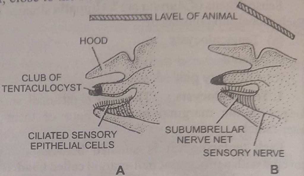
Each tentaculocyst is situated between two marginal lappets. It is covered with a process of bell-margin called the hood. It also connects the bases of two marginal lappets.
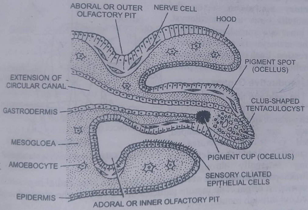
A tentaculocyst is a club-shaped structure. It receives an extension of a circular canal lined with gastrodermis. Below is base is a pad of ciliated sensory epithelial cells. In its distal part is present a mass of polygonal statolith. The epidermis is thickened and contains sensory cells at the base and sides of each tentaculocyst. Its sensory cells are connected with the subumbrella nerve net. The tentaculocysts are organs of equilibrium.
2. Ocelli: Two types of ocelli are present in each rhopalium. There are :
(i) Pigment spot ocellus consists of a patch of pigmented and sensory epidermal cells. It lies on the outer side of the tentaculocyst.
(ii) Pigment cup consisting of a cup-shaped depression lined with pigmented and sensory gastrodermal cells. It is situated on the inner side of the tentaculocyst, close to the statoliths. The sensory cells of ocelli are photoreceptors. These are connected with their respective nervous nets.
3. Olfactory pits: Two olfactory pits in the form of depressions are also developed near each tentaculocyst.
The outer or aboral olfactory depression is on the exumbrella outer to tentaculocyst. The inner one immediately internal to the statocyst is oral olfactory depression. Each is lined by sensory epithelium and is a chemoreceptor.
6. Reproductive Organs
The male and female sex organs are borne on separate umbrellas which are similar in appearance. The gonads are horse-shoe-shaped, plaited, or frilled structures of brilliant pink or reddish-violet colour shining externally through the semitransparent membrane of the umbrella. These are inter-radial in position and ectodermal in origin. The mature gametes are shed into the stomach and are passed out through the mouth.
BSc 1st Year Lower Non-chordates Aurelia Sample Model Practice Question Answer Papers