Q.3. Give a brief account of life cycle of Fasciola and explain the significance of different larval phases in its life-history.
Or
Give an account of the life-history of Fasciola hepatica Discus its economic importance.
Ans.3. Life History of Fasciola Hepatica
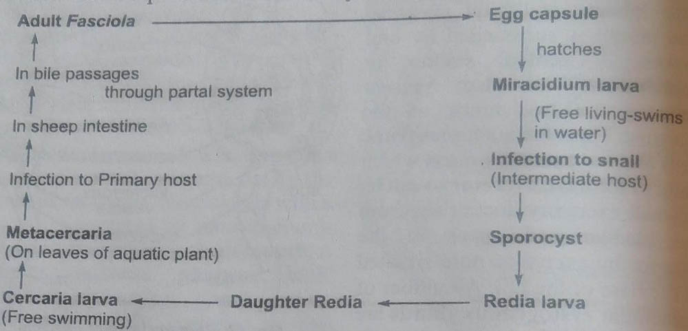
The life cycle of Fasciola hepatica is completed in two hosts. The primary or definite host is sheep or cattle, while the secondary or intermediate host is a snail of the genus Limnaea. Its life history includes a number of larval stages which propagate by asexual multiplication (polyembryony).
1. Copulation
Although Fasciola is a hermphrodite animal but cross fertilisation occurs. The two flukes copulate inside the bile duct of host’s body. The cirrus of one is inserted into the opening of laurer’s canal of the other organism and the sperm along with the parasitic fluid make their way into the Laurer’s canal, from where these move to the oviduct. Fertilisation may also occur. Sperm enters the uterus of same fluke through female genital aperture.
2. Fertilisation
The eggs are fertilised in the uterus or in ootype. The fertilised eggs are deposited with yolk cells produced by vitelline glands and are encircled by chitinous shell or egg capsule. A single fluke may produce about 2,00,000 eggs in about 11 years and 30,000 to 35,000 eggs per year.
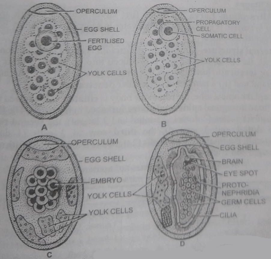
3. Eggs
The fully formed fertilised capsulated eggs are light brown, oval structures measuring about 130*150u in size. Its capsule or shell is marked off into an operculum by a distinct line. Such eggs are called operculate. Situated immediately beneath the operculum is a granular cushion. At a later stage of development, the eggs are deposited in the bile ducts of the host and carried to the intestine along with the bile and are finally passed out with faeces.
4. Segmentation and Early Development
Segmentation or cleavage starts even when the eggs are inside the uterus. The first cleavage is complete but unequal and produces (i) a small granular propagatory cell and (ii) a large somatic or ectodermal cell. Repeated divisions of the somatic cell further form ectoderm of larva. The propagatory cell further divides into two types of cells-propagative cells and somatic cells. The somatic cells after divisions form larval body structures. The propagatory cells form the germ cells. Within 9-15 days, the embryonic development is completed and a ciliated miracidium larva is formed.
5. Hatching
Further development occurs only when capsule reaches water or moist area. In contact with water, the operculum of egg capsule opens and ciliated miracidium larva hatches out. It swims in water actively.
6. Larval Stages
1. Miracidium Larva : It is the first larval stage in the life cycle of Fasciola hepatica. It swims actively in water in search of secondary host, the snail of genus, Limnaea.
External structure- Miracidium of Fasciola is about 0.07 mm long, oval or conical. Its broad anterior end is produced into an apical lobe or apical papilla or terebratorium. It bears opening of (i) a pouch-like multinucleate apical gland and (ii) a number of unicellular penetration glands.
Except the apical lobe, the rest of the body is ciliated. It is covered with 21 closely fitted hexagonal epidermal plates, arranged in five rows. The first row consists of six plates (2 dorsal, 2 ventral and 2 lateral), the second row of 6 cells (3 dorsal and 3 ventral), the third row of 3 cells (1 dorsal, 2 ventrolateral), the fourth row of 4 cells (2 right and 2 left) and the fifth row of 2 cells (1 right and 1 left) only.
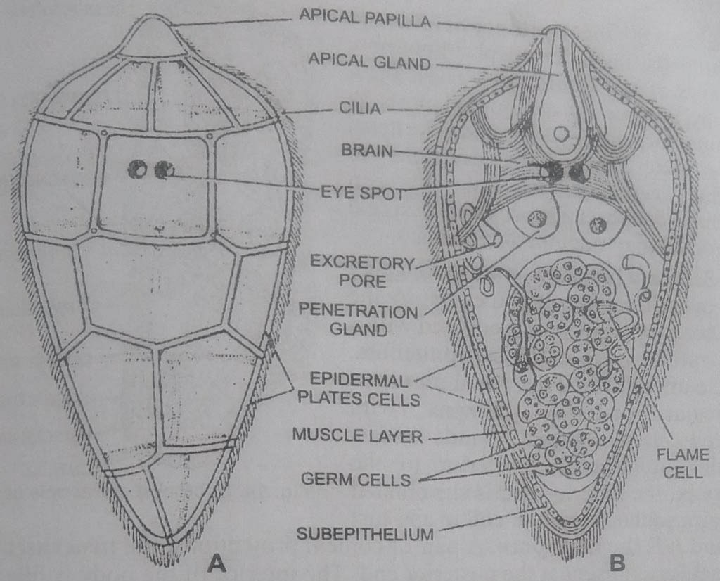
Internal structure : Below the epidermal plates is a thin layer of subepidermal musculature consisting of (i) an outer layer of circular and (ii) inner layer of longitudinal muscles. Underneath is present a subepithelial layer. The interior of larva has a pair of protonephridia and groups of germ cells. In addition, there is a pair of large pigmented eyespots, a large larval brain and a simple nervous system in the anterior part of body. There is a large apical gland and a pair of penetration glands or cephalic glands in the anterior part of body. These open on the apical lobe or cephalic cone.
7. Infection of the Secondary Host
Miracidium does not feed. It swims actively in search of its secondary host-Limnaea truncatula or Planorbis or Bulinus. In case, it fails to reach the host, it dies within 24 hours. If it finds the snail, it penetrates through snail’s soft skin or respiratory tissue. The apical lobe bores through the tissue and the tissue dissolving larval secretion of penetration glands makes a minute opening in the host tissue.(BSc Lower Non-chordates Fasciola Hepatica Question Answers)
Inside the host tissue, miracidium throws off ciliated epidermis, penetrates deeper to reach the lymph vessels or pulmonary chamber. Here it changes into second larval stage, the sporocyst larva.
1. Sporocyst larva: Miracidium loses its apical gland, penetration gland, brain and eyespots and changes into a sac-like sporocyst larva. It looks can elongated sac about 0.7 mm long. Its body wall retains all the layers of miracidium’s body wall except the ciliated epithelium. It consists of a thin cuticle, a laver of circular and longitudinal muscles. The wide interior is occupied by the protonephridia and germ cells.
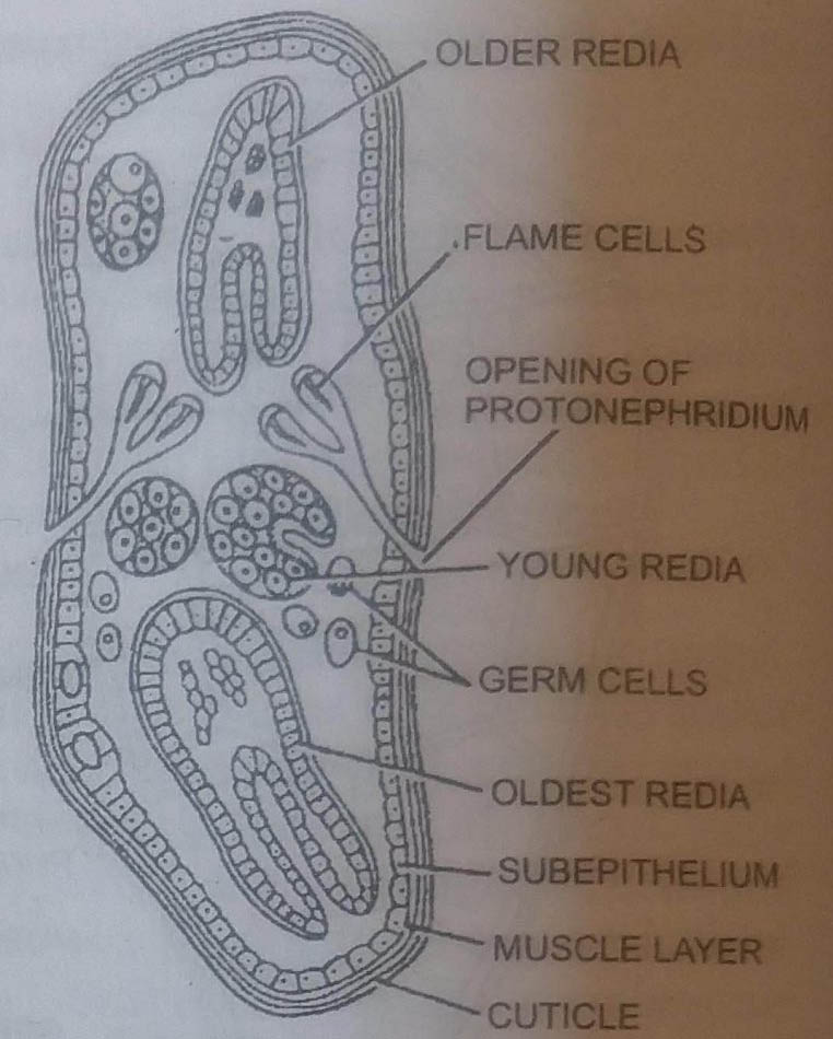
Each protonephridium now consists of two flame cells. These open on the surface by a common pore. A rudimentary gut is also found. The germ cells undergo repeated divisions to produce redia larvae, but may also produce daughter sporocysts. A single sporocyst may contain 5-18 radia.
2. Redia larva : It is an elongated and cylindrical sac. It comes out by the rupture of sporocyst. It is covered with a thin cuticle secreted by subepithelium. At the anterior end is a small mouth, a muscular suctorial pharynx with unicellular pharyngeal glands and a small gut. Slightly posterior to the pharynx, the anterior end is surrounded by a muscular band-like collar and just behind it is the birth pore. A pair of conical projections, the procruscular or lappets is present at the posterior end.
The interior of the body is filled with loose parenchyma, through which are found groups of germ cells called germ balls and highly branched flame cells. Their excretory ducts open through the paired excretory pores. The germ balls present inside redia give rise to generation of daughter rediae in summer months and produce cercaria larvae in autumn.
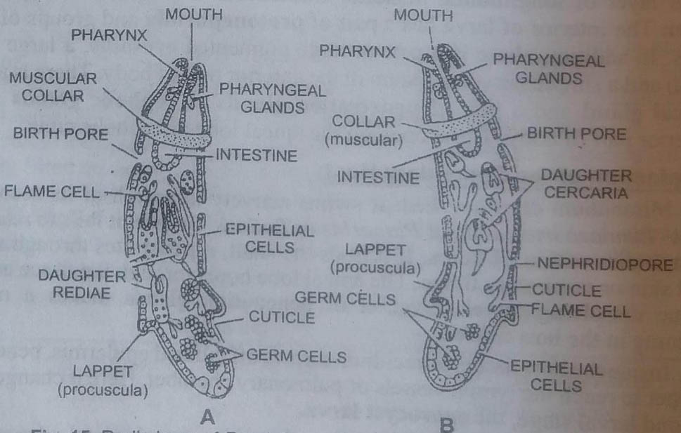
3. Cercaria larva : A fully formed cercaria larva possesses a flattened heart-shaped body with a long contractile tail. Its body surface is covered with thin cuticle with backwardly directed spines. The anterior end bears mouth, muscular pharynx, oesophagus and bifid intestine. The mouth is surrounded by oral cocker.
A ventral sucker or acetabulum is also present between the two limbs of the intestine. There are numerous flame cells and excretory ducts which unite to form the excretory vesicle. A small excretory duct arises from the bladder and opens to the exterior by excretory pore situated at the base of the tail. A number of unicellular cystogenous glands are situated below the large body wall. Their secretion forms the cyst around the larva when it is converted into metacercaria. Groups of germ cells are also present.
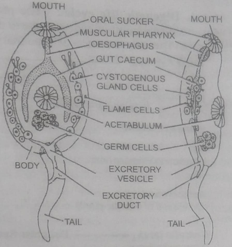
When mature, the crecaria leaves the redia through birth pore and also wriggles out of the snail body. It swims in water for some time and finally settles down on the blade of some aquatic weed. It sheds off the tail and a cyst is formed by the secretion of cystogenous glands. Thus a metacercaria is formed.
4. Metacercaria : The metacercaria is somewhat rounded with thick outer covering of cuticle in the form of cyst.
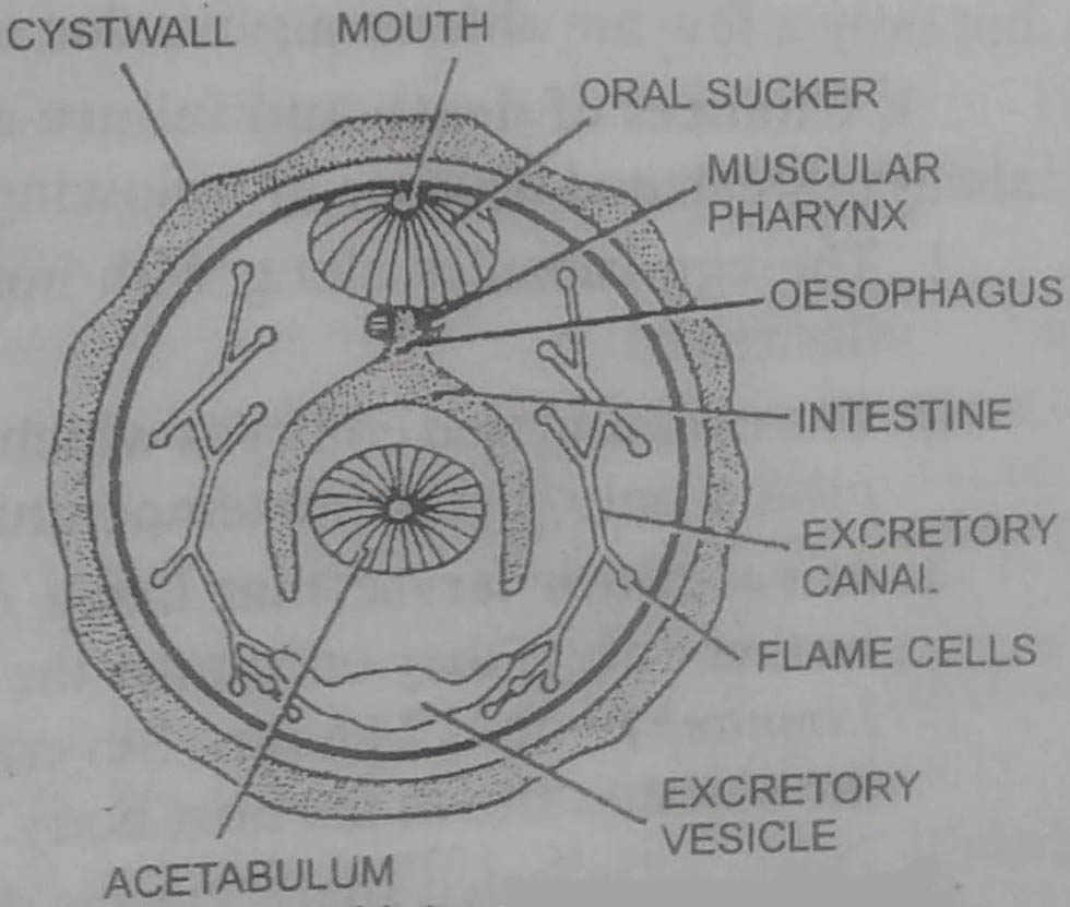
The cystogenous cells of the cercaria disappear and the flame cells increase in number.
8. Infection to the Final Host
The metacercaria enters the final or definitive host, the sheep, when it grazes on the aquatic weeds and reaches the intestine. In the intestine the cyst is dissolved by the action of digestive enzymes and young fluke comes out. It reaches the liver through the hepatic portal system and starts its existence inside the bile passage.
Significant Features in the Life-Cycle of Fasciola
Fasciola is an endoparasite. Its existence is always threatened. If it fails to enter new hosts, it will die with the death of the host. For the perpetuation of the race, the life cycle of Fasciola is digenetic and it has a number of larval stages.
1. Digenetic life cycle: The presence of two hosts in the life cycle ensures distribution of endoparasite to new hosts and helps in the perpetuation of race. But the presence of secondary host poses other problems, such as
availabity or death of parasite during transport. Therefore, there is over production and multiplication of larvae at different stages.
2. High rate of reproduction : Fasciola in its life-cycle produces more than 20,000 eggs. The eggs develop into miracidium larvae. The miracidium larvae develop into sporocysts. Each sporocyst produces 5-8 radiae, each redia larva produces 8-12 rediae in 2nd generation, each of which produces 14 to 20 cercariae. Thus each egg is capable of producing around 1000 to 2000 adults but only a few are able to survive due to high rate of mortality.
3. Chances of death and failure of life cycle : Survival of Fasciola is always threatened because of following reasons :
1. The egg capsules along with host faeces, if released on dry place are destroyed.
2. The encapsulated embryos which come out with host faeces can develop further only if suitable temperature, pH and moisture are available.
3. Miracidium larvae that hatch out of the egg capsules, can develop further only if they encounter the suitable secondary host (snail of genus Limnaea) within 24 hours. Several miracidia die midway failing to reach the suitable site in the host body.
4. Death of infected snails causes death of miracidia along with host.
5. Drying of ponds may kill miracidia and cercaria.
6. Cercaria larvae, if ingested by some vertebrate host even before these have changed into metacercaria, are digested by the gastric juice of the host.
7. Metacercaria are destroyed if ingested by host other than the primary host. If exposed to a long period of dessication, these die.
4. Polyembryony: When several embryos are produced from a single cell, it is called polyembryony. The zygote of Fasciola divides into a somatic anda propagatory cell. The cells produced from propagatory form called germcells in the larval stages. The redia, daughter redia and cercaria larval stages develop from these germ cells one after the other. According to the theory of polyembryony, the appearance of various larval forms from germ cells can be taken as simple asexual multiplication.
5. Extended metamorphosis: This has also been presumed that sexual multiplication of larval forms is an extended metamorphosis due to parasitism.
6. Heterogamy: Grobben (1882) suggested that the germ cells in different larvae were eggs that developed into next larval stage by parthenogenesis. This type of asexual multiplication is called heterogamy and various larval stages are parthenogenic forms. But this theory has been discarded.
7. Advanced larval stage: Body cavity, sense organs and locomotory organs which are lacking in the adult are present in the larvae. Thus miracidium and cercaria which are free living larvae are more advanced than the adult. The adult has degenerated in many respects to suit its parasitic mode of life.
8. Complex life cycle: The presence of several larval stages has made the life-cycle of Fasciola complicated.
9. Alternation of generations or metagenesis: It was presumed that the adult fluke represents the sexual generation, and the sporocysts, rediae and cercaria are the asexual or parthenogenetic generations. These two generations alternate with one another. Hence the life-history of Fasciola exhibits alternation of generations.
ECONOMIC IMPORTANCE OF FASCIOLA HEPATICA
The disease caused due to infection of Fasciola hepatica is called fasciolepsis. The liver fluke lives in the bile passages of liver of sheep. Its presence causes hepatitis and inflammation in the bile ducts. The calcification of bile passages results in the formation of gall stone causing damage to the liver, described as liver rot.
The liver rot may be acute or chronic. In acute liver rot the sheep becomes sluggish, feels abdominal pain with loss of weight and enlargement of liver. So the host dies in a few days. The chronic liver rot is caused by slow and successive entry of cercaria which causes anaemia and loss in weight and ultimately the watery swellings in the jaws. The unthrifty sheep dies within a few months.
Liver fluke which is parasitic in man causes liver diseases, occlusion of bile passages and anaemia. It causes body pain, cough and dysentry.
In snails, redia damages the host tissue while migrating from pulmonary sac and may cause death of snail.
Preventive Control Measures
1. Killing heavily infected sheep.
2. Destroying eggs and manure of sheep.
3. Killing snail population.
4. Man should eat thoroughly washed and properly cooked vegetables
5. Antihelminth drugs such as tetrachloro-ethane, emetine hydrochlorides phenothrazine are beneficial in treating cases of liver rot.
BSc 1st Year Lower Non-chordates Fasciola Hepatica Sample Model Practice Question Answer Papers