BSc 1st Year Lower Non-chordates Monocystis Sample Model Practice Question Answer Papers
BSc 1st Year Lower Non-chordates Monocystis Sample Model Practice Question Answer Papers: BSc is a three-year program in most universities. Some of the universities also offer BSc Honours. After getting enrolled for BSc, there are certain things you require the most to get better grades/marks in BSc. Out of those, there are BSc Study Material, BSc Sample Model Practice Mock Question Answer Papers along with BSc Previous Year Papers. At gurujistudy.com you can easily get all these study materials and notes for free. Here in this post, we are happy to provide you with BSc 1st Year Lower Non-chordates Monocystis Sample Model Practice Question Answer Papers.

Index for BSc 1st Year Lower Non-chordates Monocystis Sample Model Practice Question Answer Papers
The structure and life-history of Monocystis: Page 1
The effects of parasitism on Monocystis: Page 2
Q. 1. Give an illustrated account of the structure and life history of Monocystis.
Ans.1. Structure
Habits and Habitat
Monocystis is an extracellular parasite in the seminal vesicles of earthworms.
Shape and Size
The adult feeding and growing stage of Monocystis is known as trophozoite stage. The young trophozoite is small and ellipsoidal. It remains embedded in a group of developing spermatozoa. It appears ciliated as the tails of the dead sperms remain attached to it.
The mature trophozoite is elongated, spindle-shaped and dorsoventrally flattened worm-like creature. It is visible to the naked eye as a white thread.
Pellicle
The body is bounded by a thick, firm but elastic pellicle. It is formed of nitrogenous matter.
It may be striated or thrown into ridges and furrows or with hair-like processes. It contains contractile microtubules.
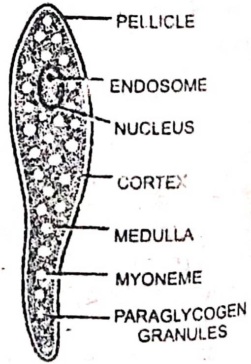
Cytoplasm
The cytoplasm is differentiated into two layers :
1. Cortex or ectoplasm: It is the outer clear laver of cytoplasm which is further differentiated into three layers :
(a) the outer layer-epimyocyte,
(b) the middle layer-sarcocyte and
(c) the inner layer, myocyte
The myocyte is traversed with longitudinal and transverse contractile fibrils, the myonemes. These help in the metabolic contraction of body and wriggling or gliding movement.
2. Medulla or endoplasm: It is the inner granular mass consiting of minute granules of paraglycogen (the reserve food), fat globules and sometimes volutin (protein).
Nutrition : Monocystis has saprozoic mode of reticulum nutrition. It secretes digeslive enzymes from its body which digests cytoplasın and developing sperms in the seminal vesicles of the host. The digested food in absorbed by osmosis through the pellick.
3. Nucleus
The nucleus is large, vesicular and ovoid or spherical. It lies towards the anterior end and is bounded by a distinct nuclear membrane. It contains one or sometimes two nucleoli, endosomes or karyosomes.
The parapodia, cilia, flagella, cell mouth etc., and food vacuole, and contractile vacuoles are absent due to parasitic mode of life.
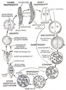
Life Cycle
Life-cycle of Monocystis is monogenetic. It is completed within one host (earthworm). The life-cycle can be distinguished into following phases :
A. Gamontogamy
It is sexual phase of life-cycle. It includes (i) pairing of gamonts, syzygy (ii) formation of gametes, gametogony and (iii) syngamy or fertilisation :
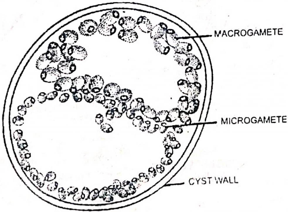
1. Syzygy: After leading a short extracellular existence in the vesicles and attaining maturity, the trophozoite changes into a spherical or oval gamont or gametocyte. These come together and closely oppose each other. These secrete a common double-layered gameto-cyst. It is hard, chitinous and protective. Its rigid and thick outer layer is known as ectocyst, while the inner thin membrane as the endocyst. The gametocystes remain closely oppressed together inside the gameto-cyst but they never fuse and are separated by a distinct wall. This type of association of the gameto-cytes is known as syzygy.
2. Gametogony: The nucleus in cach gametocyte divides by repeated divisions. The last one is reduction division while otehrs are simple mitotic. As a result a large number of haploid nuclei are formed in each gametocyte. These get arranged at the periphery and protrude out. The gametocyte now assumes appearance of a battery. Each nucleus gets surrounded by a little cytoplasm 10 form the gamete. These are liberated by the rupture of intervening wall of gametocytes.
The gametes thus formed in the two gametocytes are similar in their morphology but of two different sizes-small motile microgametes and large, less motile and somewhat rounded macrogametes.
3. Synamy: The gametes so formed move freely for some time and then pairing occurs between the gametes of two different gametocytes. Although the gametes are similar in appearance and structure but fusion always occurs between the gametes formed from the different gametocytes. The process of their fusion is known as syngamy. As a result of fusion gyzotes are formed which contain diploid number of chromosomes as found in the trophozoite.
4. Sporogony
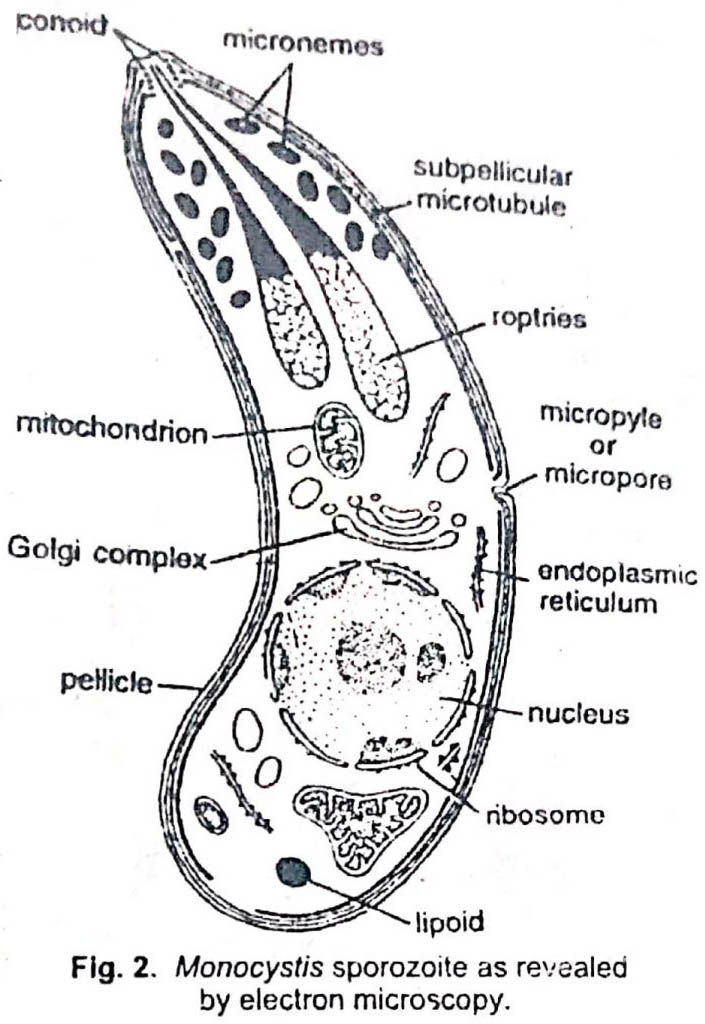
The development of the zygote occurs within the gametocyst. Each developing zygote is known as sporoblast. It becomes oval and secretes a tough, single-layered chitinous sporocyst or spore case. Thus sporoblasts change into spores. Later on, the sporocyst becomes boat-like and is termed as pseudonavicella. The nucleus inside the sporocyst undergoes three successive mitotic divisions. Simultaneously, the cytoplasm also divides. As a result eight sporozoites are formed inside each sporocyst. These are elongated and sickle-shaped.
The spores containing sporozoites are set free inside the seminal vesicles by the rupture of gametocyst. The release of sporozoites and their further development takes only inside the alimentary canal of new host.
Transmission of Spores and Infection of New Host
The transmission of spores from the infected into the fresh host occurs in the following ways :
1. During copulation: The spores of transmitted from one worm to the other during copulation, because these are seen in the sperm ducts and cocoon.
2. Due to death of host: As a result of death and decay of the host body, the spores are liberated into the soil and they make their way into the alimentary canal of other worms which feed on that mud.
3. By birds: When an infected earthworm is eaten by a bird, the spores are liberated in its alimentary canal and come out unaltered with its faeces. These along with mud enter the alimentary canal of the worm.
Inside the alimentary canal of earthworms, the spore wall gets dissolved by the digestive juices, and sporozoites are liberated. These penetrate the wall of the alimentary canal and enter seminal vesicles.
Each sporozoite enters a sperm morule and starts trophozite phase of the life cycle. (BSc 1st Year Lower Non-chordates Monocystis Sample Model Practice Question Answer Papers)
BSc 1st Year Lower Non-chordates Monocystis Sample Model Practice Question Answer Papers