Q.3. Give an account of the morphological organization and affinities of Ctenophora.
Or
Give an account general organization of Hormiphora and discuss the affinities of the phylum Ctenophora.
Ans.3.
Definition
Ctenophora is a small phylum of marine animals commonly known as walnuts or comb jellies. These are biradially symmetrical and have a transparent gelatinous body without nematocysts but with special lasso cells and with eight locomotory comb-like ciliary plates (comb plates).
External Features
Shape: The generalised ctenophore (like Pleurobrachia) is roughly spherical, ovoid, or pear-shaped in appearance.
Size: Ctenophores range in size from a few millimeters to 20 cm. Though a few species of Cestum are as long as 1 meter or more.
Colour: These are usually transparent but structures like tentacles and comb rows may be tinged with white orange or purple.
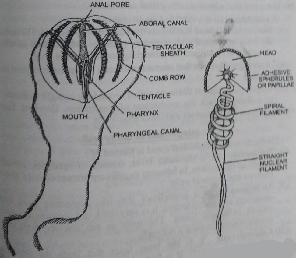
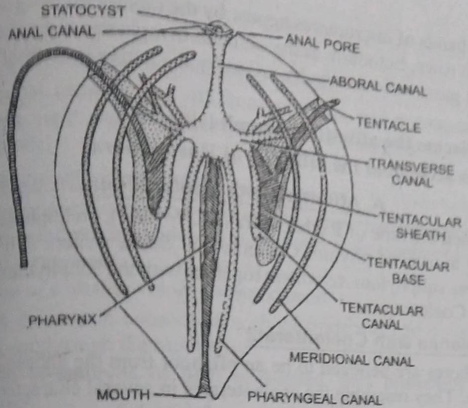
Division of body: The spherical body can be divided into two hemispheres (biradial symmetry). It can further be divided into equal sections by eight ciliated bands. The oral and aboral ends are distinct. The oral end lies on the lower side and bears a minute oral aperture or mouth. The upper end represents the aboral pole and bears an apical organ.
1. Comb plates: There are eight equally-spaced ciliated bands or paddle plates. These are arranged on the sides of the body like meridians from the aboral pole to the oral pole. These plates are known as comb rows and are characteristic of all ctenophores.
Each comb row is formed of short transverse plates of long fused cilia called comb plates. The combs in a comb row are arranged in succession one behind the other. (BSc 1st Year Lower Non-chordates Phylum Ctenophora Sample Model Practice Question Answer Papers)
The rows of ciliated bands form roughened ridges, similar to those of a walnut (hence commonly called walnuts). These help in locomotion.
2. Tentacles: In the aboral hemisphere, near the aboral end are present two long, solid, and branched tentacles. These are enclosed in a pair of deep blind pouches or the tentacular sheaths into which these can be withdrawn wholly or partially. Each tentacle bears a row of short branches or pinnae. It is formed of a mesenchymal core covered with an epidermis. It lacks nematocysts but bears peculiar adhesive cells, called colloblasts.
A colloblast cell is a modified epidermal cell. It has :
(i) Hemispherical head that lies on the surface of the epidermis. The surface of the head is covered with secretory granules that produce an adhesive secretion to be utilised for entangling and killing the prey.
(ii) A straight filament that connects the head to the mesenchymal core of the tentacle.
(iii) A spring-like spiral filament coiled around the straight filament.
The colloblasts lack the mechanism of penetration. However, Euchlore Rubra possesses nematocysts and lacks lasso cells.
Integument or Body wall
The body wall is diploblastic and is formed of:
(i) Epidermis: It is the outer layer, formed of syncytial, cuboidal columnar epithelium. In between its cells are present sensory, glandular, mucous, and pigment cells.
(ii) Mesogloea: It is a non-cellular jelly-like cementing layer between the epidermis and gastrodermis. It is very thick and contains amoebocytes, muscle cells, and fibres. The muscles are smooth, branching, and anastomosing forming a network.
(iii) Gastrodermis: It is the inner layer that lines the stomach and gastrovascular canals. It is formed of tall vacuolated and low-ciliated cells.
Digestive System or Gastrovascular System
The mouth is a slit-like aperture, situated at the lower end. It leads into a long tubular pharynx or stomodaeum. Its wall is folded or plicated and is formed of long ciliated epidermal and glandular cells. It leads into a wide stomach or infundibulum which is connected with a system of 5 gastro-vascular canals. These extend through the mesogloea in a definite arrangement up to the aboral pole.
The nervous system is of diffused type and is formed of a general subepithelial nerve plexus with multipolar ganglionic cells and neurites throughout the surface.
Reproductive System
Ctenophores are hermaphrodite. Gonads are situated in the wall of Meridional canals in the form of continuous or discontinuous bands. The gonads are derived from the endoderm. The mature gametes are discharged into the cavity of the meridional canals. These are ejected out through the mouth.
Development
Fertilisation is external. The zygote undergoes two meridional cleavages forming four blastomeres. The third cleavage is nearly vertical and results in the formation of a curved plate of 8-cells, that are arranged in two rows. These divide twice by two horizontal divisions producing each time eight small cells (micromeres) and eight large cells (macromeres).
The micromeres give rise to ectoderm and macromeres form endoderm. Micromeres undergo rapid divisions and form a sheath of small cells over macromeres which also form a sheath of cells. As a result of invagination, gastrula is formed.
Four inter-radial bands of micromeres appear by the rapidly dividing cells that give rise to comb rows. The ectoderm at the oral end invaginates to form stomodaeum from which the gastrovascular cavity arises by the active growth of endodermal cells.
BSc 1st Year Lower Non-chordates Phylum Ctenophora Sample Model Practice Question Answer Papers