Q. 3. Describe the post fertilization stages in Marchantia.
Ans.3. Zygote or fertilized egg is the first cell or unit of sporophyte of Marchantia. After fertilization the archegonium enlarges, due to which the sporophyte is continually enclosed within the archegonium, which ultimately forms a protective sheath the calyptra. In addition the surrounding gametophytic tissue and cells below the archegonium also set stimulus, divide and redivide and form a cylindrical out growth the pseudoperienth.
Thus, the mature Marchantia sporophyte has three protective coverings a calyptra, pseudoperienth and involucre.
At the time of development the zygote increase in size and fills the cavity of venter, and secretes a wall around it. The zygote first of all divides by a transverse division into two cells, the upper hypobasal and lower epibasal. Now both the cells further divide by a vertical wall forming quadrat stage.
The two upper hypobasal cells give rise to foot and seta, while epibasal half develops into capsule. The four celled zygote or embryo further divides by a vertical wall at right angles to each other the forming octant stage or 8 celled embryo.
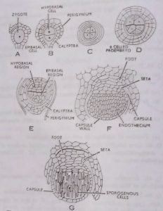
Now the embryo elongates and further divides by several irregular divisions so as to form a multicellular globular embryo. This multicellular embryo gets differentiated into foot cell, seta cells (from hypobasal region) and capsular cells from epibasal region. Now the sudden increase in length of the seta takes place by an elongation of its cells growth beyond such enveloping structure as involucre and pseudoperianth.
Cell division in development of a foot is at various angles to one another. When mature, the foot may be bulbous as in M. polymorpha. The foot serves as an organ for anchorage and for absorption of food from adjoining gametophytic tissues. However the presence of some chloroplasts shows that sporophyte is not wholly dependent upon the gametophyte for the products of photosynthesis (Bold 1918). The capsular part of embryo soon divides by periclinal division so as to form outer amphithecium and inner endothecium.
The amphithecium forms the wall of capsule, while endothecium forms archesporium which in turn forms the sporogenous tissue and spores. The archesporium divides and redivides, and forms the sporogenous tissue. The sporogenous tissue get separated to from elaters, while rest of sporogenous cells divide transversely to form vertical rows or spore mother cell or sporocytes. The spore mother cell division by a reduction division (meiosis) and followed by a simple division to form four spores from each spore mother cell. The spores are haploid in nature.
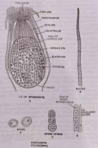
The mature sporangium is an elongated structure differentiated into definite foot, seta and capsule. The foot is a bulbous structure towards the base of archegonium it helps in absorption and anchoring. The seta is short, thick stalk connecting foot and capsule. The capsule is a spherical structure at the distal end of sporogonium. It has a sterile jacket layer of cell.
Inside the wall there are spores and elaters. The elaters are long, narrow spindle-shaped cell pointed at each end, with two spiral thickening on their walls. These elaters are hygroscopic in nature. The spore is spherical haploid structure with two layered wall-exine and intine. The exine being thick and warty. Inside the walls of spores there is a haploid nucleus and cytoplasm along with some chloroplasts. (BSc Botany Marchantia Notes Study Material)
Dispersal of Spore and Dehiscence of the Capsule
The sudden elongation of seta ruptures the calyptra and exposes the capsule from its three protective sheaths, the calyptra, the pseudoperianth and the perichaetium. Soon after ‘exposure’ to the outside atmosphere, the wall of the capsule splits longitudinally from the apex to about the middle and into an indefinite number of segments or lobes which roll back slightly thus exposing a protuberant mass of spores and elaters.
The elaters are hygroscopic; they twist during dry weather but uncoil during moist weather and thus help to loosen the spore mass and disperse the spores into the air.
The Germination of Spore and Development of Gametophyte
The spores of Marchantia remain viable for one year. Out of the four spores produced in a tetrad, usually two develop into male thalli whereas the other two produce female thalli. Thus, though the spores of Marchantia are morphologically all alike, they are of two types genetically, hence, Marchantia is an example of functional heterospory.
The spore is the first cell of the gametophyte. The spores are spherical and very small structure having a spore coat of endospore and exospore. The exospore or exine is thick and warty. While endospore is thin. Inside the wall of spore a single nucleus with granular cytoplasm.
The spores just after dispersal on getting the suitable condition start germination. The spores increases to size and its exospore rupture and endospore comes out in a form of short filaments. The filament like structure by division and redivision forms the multicellular structure and finally is get changed into dichotomously branched young gametophytic thallus of Marchantia.
BSc 1st Year Sample Model Practice Mock Test Question Answer Papers