BSc 1st Year Botany Spore Dispersal in Bryophytes Sample Model Practice Question Answer Papers
BSc 1st Year Botany Spore Dispersal in Bryophytes Sample Model Practice Question Answer Papers: BSc is a three-year program in most universities. Some of the universities also offer BSc Honours. After getting enrolled for BSc, there are certain things you require the most to get better grades/marks in BSc. Out of those, there are BSc Study Material, BSc Sample Model Practice Mock Question Answer Papers along with BSc Previous Year Papers. At gurujistudy.com you can easily get all these study materials and notes for free. Here in this post, we are happy to provide you with BSc 1st Year Botany Spore Dispersal in Bryophytes Sample Model Practice Question Answer Papers.
-
BSc 1st Year Botany Spore Dispersal in Bryophytes Sample Model Practice Question Answer Papers
Q. 1. Discuss the various methods of spore dispersal in bryophytes studied by you.
Ans.1. The spores which are the units of gametophytes produced within the sporophyte or capsule from the spore mother cell after reduction division are the minute uninucleated spherical structures. The spores represent the first stage of gametophytic or haploid generation. The mechanism of the dispersal of these spores greatly varies in different genera of Bryophyta depending upon the structure and position of the sporophyte.
The important methods of spore dispersal are as follows:
(i) In Riccia there is no special mechanism for spore dispersal. In this case the sporophyte is a globular structure which remains completely embedded in the tissue of the gametophyte. Just before maturation of spores, the wall of capsule disintegrates. The outer layer of calyptra and adjacent thallus decays and spores are left in the soil, which are later dispersed to longer distance by the wind.
(ii) In Marchantia, the spore dispersal takes place by the help of hygroscopic movement of elaters. In this case the sporophyte is differentiated into foot, seta and capsule. In the capsule are present numerous spores and elater.
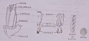
The elaters are with annular thickening and are sterile. In this case the wall of capsule splits longitudinally into a number of lobes, elaters also help in the dispersal of spores. The wall of capsule splits open into 4 to 6 teeth like structure exposing elaters and spores. The hygroscopic elaters loosen the spores, which are carried by the wind.
(iii) In Pellia, the mechanism of spore dispersal is nearly the same as in Marchantia with the only difference that in the case capsule splits into four longitudinal valves only.
(iv) In Anthoceros, the spores dispersal takes place by the help of pseudoelaters. The sporophyte, in this case is highly advanced and is differentiated into foot, rudimentary seta and capsule. The capsule has a columella, overarching spores and pseudoelaters and the wall of capsule. The wall of capsule in this case splits into 1-4 valves, which remain united of the apex. Pseudoelater is hygroscopic. In nature, they help in the dispersal of spores. Dehiscence is dependent on the loss of water.
(v) In Sphagnum, the spore dispersal takes place as a result of air pressure which is developed inside the spore sac in dry conditions. The sporophyte of Sphagnum is differentiated into foot, seta and capsule. According to Nawaschin (1897), capsule on maturation reptures violently. This results from the drying up of columella which is replaced by air. This is compressed by the contraction of the wall of the capsule on drying. This exerts considerable pressure on the enclosure layer. As results of pressure, the air forces the operculum of the capsule below up, which is thrown away to a distance of several feet and the spores disperse.
(vi) In Funaria (moss) the spores dispersed by air and gradual activity of peristomial teeth. In this case the sporophyte is differentiated into foot, seta and capsule, and show a great advance in the mechanism of spore dispersal. As the capsule matures, a number of changes takes place. The columella other internal tissue break down leaving the spore sac filled with lose mass of spores. The thin walled cells holding the operculum also breaks down and it becomes detached and thus expose the double series of teeth of peristome.
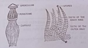
The peristome teeth are rough and are very sensitive to moisture. Each teeth of the outer peristomes is formed of two layers one outer and one inner. The outer layer is very sensitive to moisture but the inner one not so sensitive. The outer layer of the outer peristomial teeth is hygroscopic but the change takes place in the inner layer of teeth.
The inner peristomial layer shows no hygroscopic movement. When the outer peristomial teeth are wet, their outer layers increase in height and thus bend into the cavity of the sporogonium which dry again strenghten with a jerk and thus lift out some of the spores.
The inner peristomal layer does not show such movement but act as a sieve allowing dispersal of only a few spores at a time.
It being slender and hygroscopic by the combined efforts of the peristome and set the spores are liberated from the capsule. The spores after coming out from the capsule are disseminated by air. (BSc 1st Year Botany Spore Dispersal in Bryophytes Sample Model Practice Question Answer Papers)
BSc 1st Year Botany Spore Dispersal in Bryophytes Sample Model Practice Question Answer Papers
BSc 1st Year Sample Model Practice Mock Test Question Answer Papers