BSc 1st Year Botany Sporophyte in Bryophytes Sample Model Practice Question Answer Papers
BSc 1st Year Botany Sporophyte in Bryophytes Sample Model Practice Question Answer Papers: BSc is a three-year program in most universities. Some of the universities also offer BSc Honours. After getting enrolled for BSc, there are certain things you require the most to get better grades/marks in BSc. Out of those, there are BSc Study Material, BSc Sample Model Practice Mock Question Answer Papers along with BSc Previous Year Papers. At gurujistudy.com you can easily get all these study materials and notes for free. Here in this post, we are happy to provide you with BSc 1st Year Botany Evolution of Sporophyte in Bryophytes Sample Model Practice Question Answer Papers.

- BSc 1st Year Botany Sporophyte in Bryophytes Sample Model Practice Question Answer Papers
Q. 1. Explain the progressive development of Sporophytes in Bryophytes.
Ans. The sporophyte in bryophytes develops from zygote. The sporophyte of bryophytes usually lacks lateral appendages and is incapable of self-nutrition. It obtains its nutrition whole or partially from the parent gametophytes to which it remains organically attached throughout its life.
The chief function of sporophytes in bryophytes is the production and dispersal of spores. In form, it varies from only a spherical spore-producing capsule as in Riccia to an elaborate object differentiated into foot seta and capsule as in Mosses. The function of the foot is anchoring and food absorbing, the seta helps in conduction.
Usually, the series starts with the simple sporophyte of Riccia and runs through that of Marchantia, Pellia, Anthoceros, and Funaria (Moss).
Regarding the evolution of sporophytes in Bryophytes two different lines of evolution have been suggested by different workers:
(1) The progressive evolution Or Theory of sterilization.
(2) Theory of progressive evolution Or Reduction theory.
(I) Theory of sterilization
Bower (1890) said that there is a progressive elaboration and on in the structure of the sporophytes. The fundamental principle upon formulating his argument as the “progressive sterilization of potentially fertile cells of sporogenous tissue”.
He tried to explain the evolution of sporophytes in the bryophytes on the basis of progressive elaboration. This theory of Bower was further supported by Caver (1910) and Campbell (1918-40). According to this theory, Riccia sporophyte is the most primitive, and the sporophytes of Funaria are most evolved as follows:
1. Riccia sporophyte
In Riccia the zygote by divisions becomes a multicellular structure. At this stage the embryo divide by pericilinal wall into outer amphithecium and inner endothecium. In this case, the endothecium is the first cell generation (archesporium) of the sporogenous tissue, or we may say that in Riccia archesporium is formed from the entire endothecium and is centrally situated. The amphithecium forms a single-layered jacket. As a result of repeated divisions, the archesporium transforms into sporogenous cells which function as spore mother cells. (BSc 1st Year Botany Sporophyte in Bryophytes Sample Model Practice Question Answer Papers)
Most of the spore mother cells from the spores are haploid after reduction division except a few cells as nurse cells which degenerate afterward and serve as nutritive tissue. (Botany Sporophyte in Bryophytes Sample Question Answer Papers)
The sporophyte in the case of Riccia represents the simplest structure among bryophytes as it has only a capsule with a jacket and spores. There is no foot, seta, and no sterilization of sporogenous tissue in the capsule.
2. Marchantia Sporophyte
In Marchantia, the zygote divides by successive division and is differentiated into four superimposed tiers of four cells each. It divides periclinally into amphithecium and endothecium and in Marchantia, the archesporium develops from endothecium which is centrally situated. The archesporium divides and redivides and forms a massive sporogenous tissue.
The sporogenous cells elongate, some remaining undivided to form elaters, and others divide transversely by five successive divisions thus sporogenous cells forms a transverse row of 32 spore mother cell. The spore mother cell divides by meiosis and forms four haploid spores In this way each sporogenous cell either gives one elater of 28 spores.
The foot is a bulbous structure towards the base of the archegonium, it helps in absorption and anchoring. The seta is a short, thick stalk connecting the foot and capsule. The capsule is a spherical structure at the distal end of the sporogonium. It has a sterile jacket layer of cells.
Inside the wall, there are spores and elaters. The elaters are long, narrow, spindle-shaped cells pointed at each end, and with two spiral thickening on their walls. These elaters are hygroscopic in nature. The exine is thick and warty. Inside the walls of the spore, there is a haploid nucleus and one cytoplasm along with some chloroplast. (Botany Sporophyte in Bryophytes Sample Question Answer Papers)
The capsule of Marchantia has specialized both as a spore-producing and spore-distributing body. It illustrates a further step in the progressive sterilization of sporogenous tissue.
3. Pellia sporophyte
In Pellia, the archesporium formed from the entire endothecium. The sporogenous cells in the centre form elaterophore and rest spore mother cells. Archesporium remains surrounded by two or more layers of the jacket.
Thus, a fully mature capsule consists of a foot, seta, and capsule. The capsule is spherical in outline. Inside the capsule, there are present spores elaters, and elaterophore. When the capsule is fully mature, the seta suddenly elongates and the capsule comes out in the air.
A maturity the wall of the capsule splits longitudinally but into four valves only. The elaters and elaterophore help in the dispersal of spores.
4. Anthoceros sporophytes
This illustrates a further step of progressive sterilization of potentially fertile tissue. In this case, the entire endothecium remains sterile and forms a central region of the columella. (BSc 1st Year Botany Sporophyte in Bryophytes Sample Model Practice Question Answer Papers)
In Anthoceros the zygote divides to form the octant. The upper four cells of the octant give rise to the capsule. The cells divide once or twice transversely and then the periclinally differentiating into amphithecium and endothecium.
The archesporium in Anthoceros develops from amphithecium instead of endothecium. The endothecium forms the central sterile columella. The archesporium is superficial in position and over arches the apex of columella. The sporogenous cells from the spore mother cells and Pseudoelaters. The spore mother cells as a result of reduction division form spores.
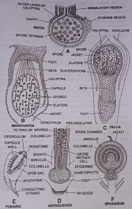
The mature sporophyte or sporogonium is an exact cylindrical structure, consisting of a bulbous foot and smooth slender, erect cylindrical capsule which may project 2 or 3 centimeters but sometimes up to 15 cm above the surface of the gametophytic thallus. (Botany Sporophyte in Bryophytes Sample Question Answer Papers)
The foot absorbs food from gametophytes. The capsule is made up of several tissues. It has a central column of sterile cells the columella, columella surrounded by the sporogenous tissue which consists of spores and elaters. The wall of the capsule is 2 to 5 layered, with an outer layer of the epidermis that has stomata.
5. Funaria sporophyte
In Funaria the zygote as a result of division form eight outer cells encircling the four inner cells. The outer cells form the amphithecium and the inner cells form the endothecium. The endothecium cells divide by periclinal divisions to form outer endothecium and inner endothecium cells. (Botany Sporophyte in Bryophytes Sample Question Answer Papers)
The inner endothecium cells give rise to columella while the outer endothecium cells form the archesporium.
Archesporium is superficial in position and surrounds the columella from all sides. The entire archesporium or sporogenous cells form the spore mother cells. The spore mother cells divide by meiosis and form haploid spores. No elaters are formed in Funaria. (BSc 1st Year Botany Sporophyte in Bryophytes Sample Model Practice Question Answer Papers)
The mature capsule when seen in the longitudinal section consists of a cap-like operculum at the top. Below the operculum, peristomes with peristomial teeth are present. (Botany Sporophyte in Bryophytes Sample Question Answer Papers)
There is present a well-developed central columella surrounded by a spore sac. Adjacent to the spore sac the air spaces are present.
The wall of the capsule is multilayered. The lower part of the capsule or apophysis has conducting strand and the epidermis of this region possesses the stomata. (BSc 1st Year Botany Sporophyte in Bryophytes Sample Model Practice Question Answer Papers)
Thus, from the above discussion, it is clear that Bower’s theory of sterilization offers a possible explanation. The theory has been accepted by most Botanists in the present day. (BSc 1st Year Botany Sporophyte in Bryophytes Sample Model Practice Question Answer Papers)
(II) Reduction theory
According to this theory of evolution, the sporophyte has a downward direction. The main supporters of this theory are Kashyap (1919), Chruck (1919), Goebel (1930), and Evans (1939). They believe and an example of retrogressive evolution. (Botany Sporophyte in Bryophytes Sample Question Answer Papers)
The reduction is accompanied by a simplification of the structure of sporophyte in the series. (Botany Sporophyte in Bryophytes Sample Question Answer Papers)
According to the view, the structure of the sporophyte of Riccia will be considered as highly evolved through reduced as a result of progressive simplification. (BSc 1st Year Botany Sporophyte in Bryophytes Sample Model Practice Question Answer Papers)
Botany Sporophyte in Bryophytes Sample Question Answer Papers
BSc 1st Year Botany Sporophyte in Bryophytes Sample Model Practice Question Answer Papers
BSc 1st Year Botany Sporophyte in Bryophytes Sample Model Practice Question Answer Papers
BSc 1st Year Sample Model Practice Mock Test Question Answer Papers