BSc 1st Year Botany Xanthophyla Sample Model Practice Question Answer Papers
BSc 1st Year Botany Xanthophyla Sample Model Practice Question Answer Papers: BSc is a three-year program in most universities. Some of the universities also offer BSc Honours. After getting enrolled for BSc, there are certain things you require the most to get better grades/marks in BSc. Out of those, there are BSc Study Material, BSc Sample Model Practice Mock Question Answer Papers along with BSc Previous Year Papers. At gurujistudy.com you can easily get all these study materials and notes for free. Here in this post, we are happy to provide you with BSc 1st Year Botany Xanthophyla Sample Model Practice Question Answer Papers.
BSc 1st Year Botany Xanthophyla Sample Model Practice Question Answer Papers
Q.1. Describe the systematic position, structure, and reproduction of Vaucheria.
Ans.1. The systematic position of Vaucheria has been the subject of debate for more than eight decades. A number of botanists had been placing Vaucheriaceae under the order Siphonales (class Chlorophyceae) following Fritsch. This was because the zoospores bear equal cilia (Isokonate) and have monogamous type of sexual reproduction.
The genus was later on shifted to Xanthophyceae. The suggestion, in fact, for its placement under Xanthophyceae had first come from Boglean (1900) followed by Blackman and Tinelsey (1900) because of the heterokont nature of antherozoids and oil as reserve food material.
Affinities of Vaucheria
(A) With members of Xanthophyceae-
(i) Siphonaceous, acellular organization of the thallus,
(ii) Photosynthetic pigments,
(iii) Discoid chloroplasts,
(iv) Cell wall composition,
(v) Flagellar morphology of motile cells,
(vi) Principal food reserves.
(B) With green algae (Chlorophyceae)
(i) Multinucleate aseptate thallus.
(ii) Oogamous sexual reproduction.
(C) With Oomycetes (Fungi)
(1) Coenocytic nature of the thallus,
(ii) Composition of the cell wall.
(iii) Presence of oogamy in the sexual phase.
The presence of whiplash and tinsel type of flagella in the motile stages of Vaucheria indicates that lower fungi have been derived from Vaucheria-like ancestors. (BSc 1st Year Botany Xanthophyla Sample Model Practice Question Answer Papers)
However, the systematic position of Vaucheria is still an open question.
Occurrence
Vaucheria occurs on damp grounds or in such places where water trickles down. It includes about 54 species. A few species of Vaucheria may be marine. The common Indian species of Vaucheria are V. sessilis, V. geminata. V. uncinata and are found covering large areas of soil from December to February.
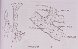
Structure
The plant body is filamentous. The filaments are branched and unseptate. The terrestrial species remain attached to the substratum by a rhizoidal basal portion or hapteron which may be brown or white in colour and is devoid of chloroplasts. The cell wall is relatively thin and consists of cellulose and pectin.
Just within the wall is a thin and thick continuous layer of cytoplasm. It contains numerous circular or elliptical chloroplasts arranged in an outer layer. Internal to the chloroplast are innumerable, minute, nuclei arranged in an inner layer. The minute droplets of oil are also found in the cytoplasm. The starch and pyrenoid is completely lacking. The cell or filament increases in length by elongation of the terminal portion of the filament.
Reproduction
Vaucheria may reproduce vegetatively, asexually, and sexually.
(1) Vegetative reproduction is secured through fragmentation.
(2) Asexual reproduction.
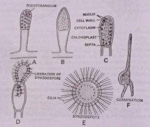
(A) By compound multiflagellate or Synzoospores formation
There are produced single in club-shaped zoosporangium. The zoosporangium is formed by a club-shaped swelling at the distal end of the filament which migrates numerous nuclei and chloroplasts. Then transverse wall cuts off the sporangium from the tip of the branch. The nuclei and chloroplast, within the sporangium reverse position, so that nuclei come to the linear periphery. The protoplast contracts slightly and develops a pair of cilia external to each nucleus. After the zoospore is fully developed, the tips of the sporangial wall soften to form a pore.
The zoospore squeezes its way through the pore and then swims freely in the water. Sometimes, the part protruded becomes separated from that still left in the sporangium and two zoospores are formed instead of one. The compound zoospores or synzoospores or coenozoospores, after the swimming period, comes or rest withdraws their cilia, and secretes a wall. Germination takes place by forming one or more germ tubes one of which forms the hold fast and others develop into the main plant.
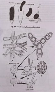
(B) By Aplanospores
Aplanospore formation normally takes place in terrestrial species with the plant is exposed to drought conditions. Single non flagellated aplanospore is formed in an aplanosporangia. The aplanosporangia develop as swellings either on the main of lateral branches. When mature aplanospore drop out through a performation in the wall, they germinate into a new filament.
(C) By Hypnospore and cyst or Akinetes
Sometimes under unfavorable periods, the coenocytic protoplast of the filament divides into numerous short segments. Each segment secretes a thick wall around it. Their protoplast becomes laden with oil.
These resting spores are called the hypnospores and this stage is known as the gongrasira stage. On the return of favorable conditions, each resting spore either directly grows into a new filament of its contents or divides into a number of thin-walled cysts. (BSc 1st Year Botany Xanthophyla Sample Model Practice Question Answer Papers)
At the time of germination the protoplast of the cysts escapes through an opening in the cyst wall. It moves about for some time in an ameoboid manner. Eventually it comes to rest assumes a spherical forms. It enclose itself by a cell wall and develops directly into new filament.
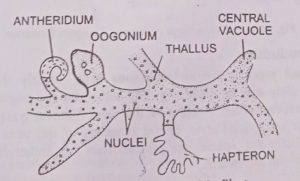
Sexual reproduction
Sexual reproduction is oogamous type. Mostly the plants are monoecious and rarely may be dioecious. The antheridia and oogonia are borne close to one another either together on the same filament lying side by side (V. sessilis) or on special short lateral branches of the same filament (V. geminata). Mostly the species are protandrous i.e., antheridium develops first and oogonium afterward. (BSc 1st Year Botany Xanthophyla Sample Model Practice Question Answer Papers)
The antheridia develop on the lateral branches at their ends shortly before the formation of oogonia. The portion of the thallus giving rise to antheridium possesses an abundance of cytoplasm chloroplasts and nuclei. The slender hook like antheridium develops having a pore at its distal end. A septum develops just beneath the curved portion. The nuclei divide again and again mitotically and around each nucleus cytoplasm is deposited and each gets metamorphosed in a biflagellate antherozoid. These antherozoids are liberated through an apical round opening of antheridium.
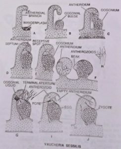
The oogonium develops as a swelling filled with nuclei, oil, and chloroplast. Usually, one large nucleus is left inside. A septum develops at the base of the oogonium. Later, a beak develops at one side, of the oogonium. A pore is formed through which antherozoids enter into oogonium.
Fertilization
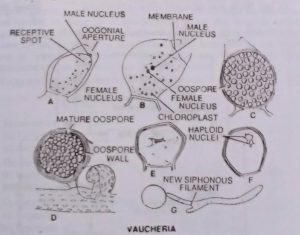
The several antherozoids on liberation try to enter the oogonium but one of them penetrates the egg. The fusion results in the formation of a zygote or oospore.
Germination of Zygote
The zygote secretes a thick wall with three to seven layers and its protoplasm becomes densely filled with oil. It changes from dark green to red or dark brown. After the long resting period, on the approach of the favorable period, the zygote nucleus divides first meiotically and then mitotically, outer walls rupture, and the inner wall forms a tube-like structure, protoplasmic contents get migrated in it and a new thallus is formed.
BSc 1st Year Botany Xanthophyla Sample Model Practice Question Answer Papers
BSc 1st Year Botany Xanthophyla Sample Model Practice Question Answer Papers
BSc 1st Year Sample Model Practice Mock Test Question Answer Papers