BSc 1st Year Lower Non-chordates Paramecium Sample Model Practice Question Answer Papers
BSc 1st Year Lower Non-chordates Paramecium Sample Model Practice Question Answer Papers: BSc is a three-year program in most universities. Some of the universities also offer BSc Honours. After getting enrolled for BSc, there are certain things you require the most to get better grades/marks in BSc. Out of those, there are BSc Study Material, BSc Sample Model Practice Mock Question Answer Papers along with BSc Previous Year Papers. At gurujistudy.com you can easily get all these study materials and notes for free. Here in this post, we are happy to provide you with BSc 1st Year Lower Non-chordates Paramecium Sample Model Practice Question Answer Papers.

Q.1. Describe the structure of Paramecium.
Ans.1. 1. Size and shape: Paramecium is unicellular microscopic organism. Its size varies in different species being 170u – 290 u in P. caudatum and 120 – 250 in P. aurelia.
Paramecium (Gr. Paramekos or parameces, oblong+L, caudata, tail) is an elongated, slipper-shaped animal and is commonly referred as slipper animalcule. Its body is asymmetrical with flat oral or ventral and a convex aboral or dorsal surface. The anterior end is rounded and the posterior end is thick and cone- shaped.
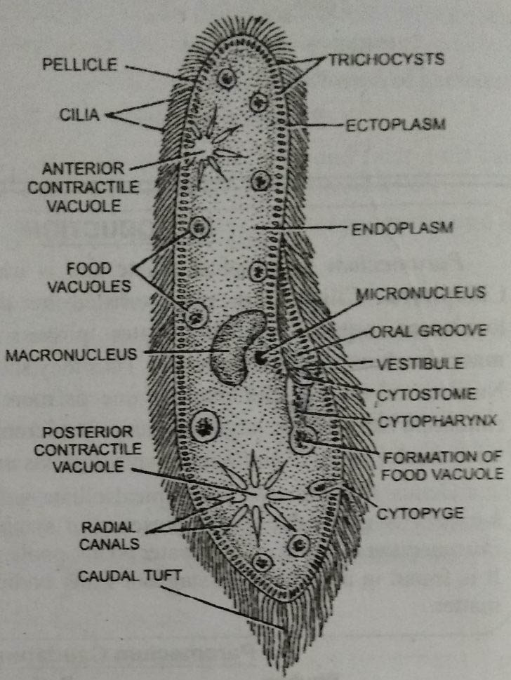
The structure is more complicated due to the development of certain organelles and can be described under the following heads :
2. Pellicle : The body is covered by a thin, firm but elastic pellicle. It gives a definite body form to the organism. The pellicle is divided into polygonal or hexagonal depressions with raised margins. A single cilium projects out from the centre of each hexagonal area. The polygonal areas correspond to regular series of cavities, the alveoli, from which cilia emerge. The anterior and posterior margins of hexagonal areas bear the openings of trichocysts.
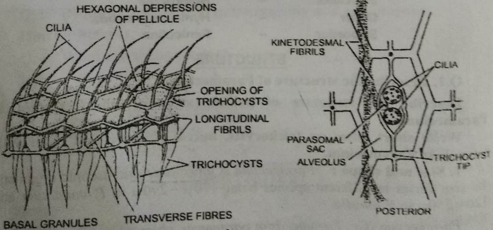
Pellicle consists of three membranes. The outer or surface membrane is continuous with the membrane surrounding the cilia. Beneath the outer membrane are closely packed alveoli. These are greatly flattened. The outer and inner membranes of the alveoli thus from the middle and inner membranes of the pellicle.
3. Cilia : The entire body surface is covered by a uniform covering of hair-like protoplasmic processes, the cilia. These emerge out from the centre of alveoli. All are of equal size except for a few at the extreme posterior end which are longer and form the caudal tuft.
Ultrastructure of cilia : Each cilium consists of fluid matrix surrounded by a membranous sheath. The membranous sheath is continuous with the outer membrane of the pellicle. Within the matrix are nine peripheral longitudinal fibres and two central longitudinal fibres. Each fibre is formed of two subfibres, one of which carries a double row of short arms all running in the same direction.
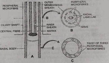
The central fibres are single and are enclosed within an inner membranous sheath. Nine very delicate accessory or radial fibrils lie between the central and peripheral fibres.
The cilia in the oral groove show variation in size and arrangement. These form following structures:
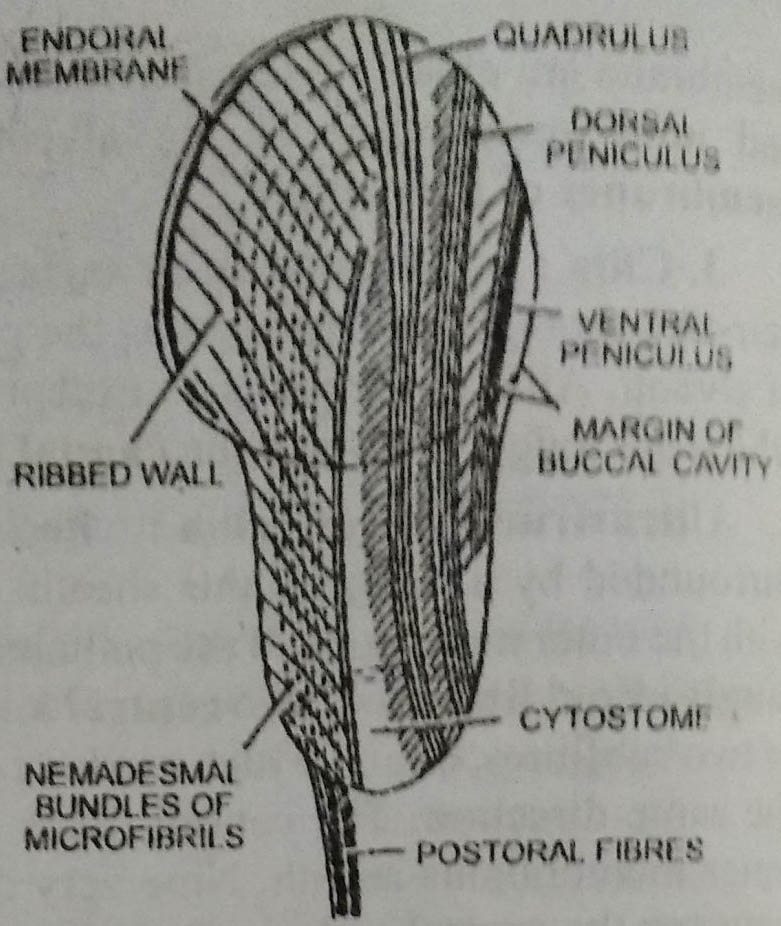
- Endoral membrane: Special cilia in the buccal cavity fuse in crescentic manner to form endoral membrane. It runs transversely and lies at the junction of vestibule and buccal cavity.
- Penniculi: There are two peniculi located in the left wall of buccal cavity. Each consists of four rows of cilia. The dorsal penniculus is longer and crosses over to the right wall at the cytostome. It terminates on the right wall of cytopharynx. The ventral peniculus is short and ends at the cytostome. The cilia of dorsal and ventral peniculi beat in opposite directions.
- Quadrulus: It is also formed of four rows of cilia. It runs along the dorsal wall of buccal cavity, crosses over to right near the cytostome and ends near the termination of dorsal peniculus,
Internal Structure
1. Cytoplasm
It is distinguished into two regions: (1) ectoplasm and (2) endoplasm
Ectoplasm or cortex: It is a narrow, peripheral, dense zone. It includes infraciliary system and trichocysts.
1. Infraciliary system: It consists of basal bodies (kinetosome) and kinetodesmata located just beneath the alveoli in the ectoplasm.
(a) Basal bodies or kinetosome: The base of each cilium is connected with a tubular basal body or kinetosome.
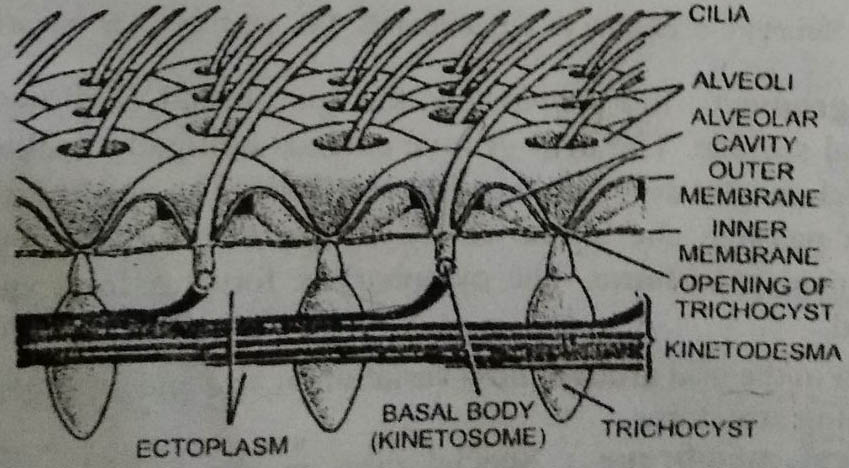
(b) Kinetodesmata: From the basal body of each cilium arises a single fibril of kinetosome. It runs anteriorly tapering along its course. It joins other fibrils of the posterior kinetosome of the same row forming a bundle of fibrils called the kinetodesma. The individual fibrils do not run anteriorly farther than the five basal bodies. Thus the number of fibrils in each kinetodesma remains five. The kinetodesma and the row of kinetosomes associated with form a structural unit called a kinety. All the kineties together form infraciliary system. This coordinates beating of cilia. Infraciliation plays an important role in the morphogenesis of Paramecium and other ciliates.
The basal granules are also said to be connected by some other types of fibrils, the myonemes and neuronemes. These are highly contractile and co-ordinate the movement of cilia. All the myonemes converge to form a darkly stained bilobed body, the motorium, situated near the cytopharynx. The basal granules, myonemes and motorium constitute the neuromotor system. It is said to provide a conducting and co-ordinating mechanism like the nervous system of higher animals. But electron microsocopy does not reveal the presence of any neuromotor system.
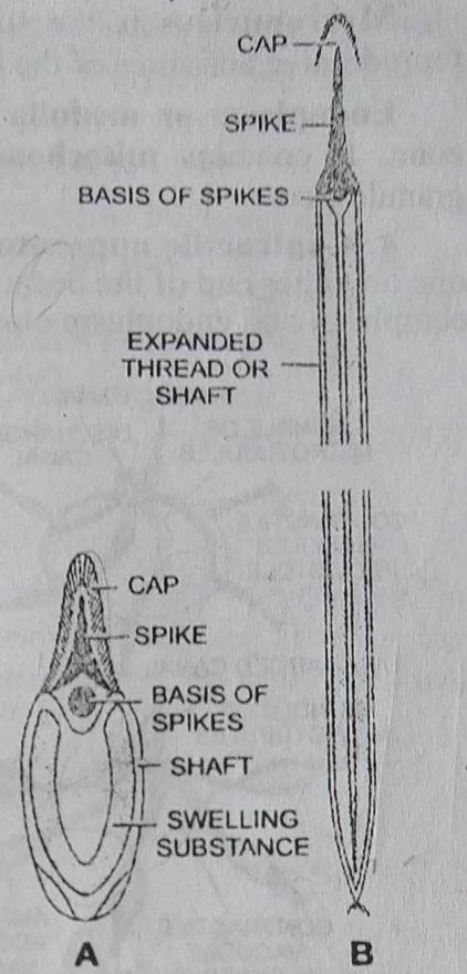
2. Trichocysts: These are rod-like or spindle- shaped. These lie embedded in the ectoplasm alternating with basal bodies and perpendicular to the body surface. Each trichocyst consists of an elongated shaft and a terminal spike or barb covered with a cap. The shaft is filled with a dense fluid having a swelling substance with a fibrous protein.
When irritated by external stimulus the trichocysts are discharged as long stick threads. The discharged trichocyst have a long striated shaft measuring upto 40u in length.
Once discharged, the trichocysts cannot be withdrawn. They are replaced by new ones. New trichocysts arise from kinetosomes of cilia and then migrate to their respective places.
The trichocysts are believed to be the organelles of defence.
3. Nuclear apparatus: It consists of a large bean-shaped macro or meganucleus situated near the cytostome and a small rounded micronucleus lodged in the depression of meganucleus.
Macronucleus is the somatic or vegetative nucleus and controls the reproductive activities of the organism.
Endoplasm or medulla: It is the large, central granular, semi-fluid zone. It contains mitochondria, ribosomes, Golgi bodies, reserve food granules, etc.
4. Contractile apparatus: There are two large contractile vacuoles, one on either end of the body. Their position is fixed and they lie between the ectoplasm and endoplasm close to dorsal surface.
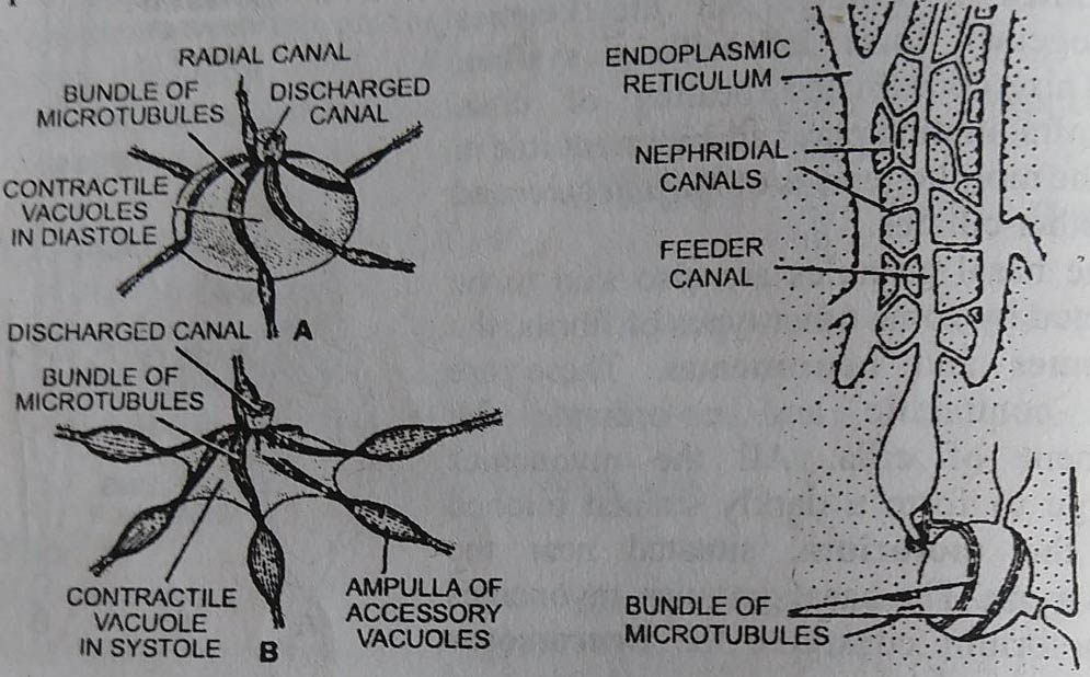
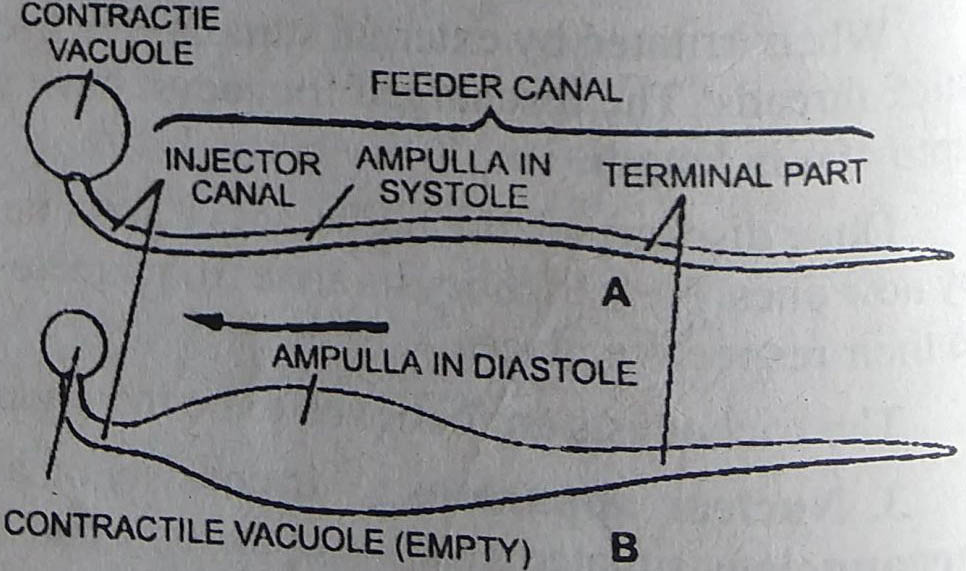
The contractile vacuole opens through a discharge canal in the pellicle on dorsal side. Each contractile vacuole is surrounded by 6-10 elongated radiating canals, which are also known as feeding canals. Each feeding canal is differentiated into :
(a) injector canal which opens into the vacuole
(b) ampulla that collapses when empty, and
(c) terminal part extends in the cytoplasm. The distal end of terminal part is associated with the nephridial canals.
The nephridial canals are connected with endoplasmic reticulum.
5. Food vacuoles: These are roughly spherical, non-contractile bodies varying in size and
number lying in the endoplasm. These contain ingested food particles.
6. Cytopyge or cyto-proct: It lies on the ventral side of the body a little behind the cytostome or mouth. It is visible only when the undigested 100 particles are eliminated through it.
BSc 1st Year Lower Non-chordates Paramecium Sample Model Practice Question Answer Papers