BSc 1st Year Botany Eukaryotic Algal Cell Sample Model Practice Question Answer Papers
BSc 1st Year Botany Eukaryotic Algal Cell Sample Model Practice Question Answer Papers: BSc is a three-year program in most universities. Some of the universities also offer BSc Honours. After getting enrolled for BSc, there are certain things you require the most to get better grades/marks in BSc. Out of those, there are BSc Study Material, BSc Sample Model Practice Mock Question Answer Papers along with BSc Previous Year Papers. At gurujistudy.com you can easily get all these study materials and notes for free. Here in this post, we are happy to provide you with BSc 1st Year Botany Eukaryotic Cell Sample Model Practice Question Answer Papers.

Index for BSc 1st Year Botany Eukaryotic Algal Cell Sample Model Practice Question Answer Papers
Structure of Eukaryotic Algal Cell: Page 1
Structure of Cyanobacterial Cell: Page 2
Q. 1. Describe the ultrastructure of the Eukaryotic Algal cell.
OR
Describe the ultrastructure of the Chlamydomonas cell.
Ans. Chlamydomonas represent, a Eukaryotic algal cell. The Chlamydomonas cell have
(a) Cell Wall
(b) Cytoplasm including cell organelles
(c) Chloroplast
(a) Cell wall: The cell wall of Chlamydomonas shows that the cell wall consists of 2-3 layers but recent investigations made by Robert et. al. (1972), Hills (1973), and Hills et. al. (1973) in C. reinhardtii indicate that the cell wall is made up of 7 layers out of which W7 is formed by the adsorption of culture medium.
Next to the cell wall, lies another membrane called Plasmalemma. Plasmalemma consists of two membranes that remain separated by an opaque zone. It is differentially permeable.
(b) Cytoplasm including cell organelles
(i) Flagella: The tip of each glagellum is narrow because of the reduction in the diameter of the central structure of the axoneme and the absence of a sheath. Internally each flagellum shows a typical 9 + 2 arrangement of the fibrillar components. The 9 peripheral pairs (doublet fibrils) surround the central 2 (singlet) fibrils. The flagella of C. reinhardtii produce through the cell wall via short tunnel-like openings that are lined with fibres arranged in parallel array. These cylindrical collections of fibres presumably permit free movement of flagella within the cell wall.
(ii) Eye spot: The eyespot is made up of 2-3, parallel rows of droplets called granules that contain carotenoids. Each granule is about 75 mm in diameter. Rows of granules are separated by lamellae of the chloroplast. The eye spot is a part of the chloroplast and remains embedded in it. It has got no association with any of the flagella.
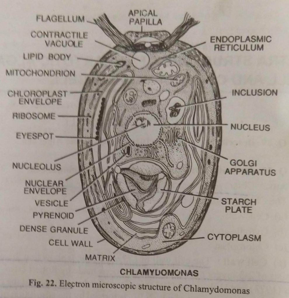
(iii) Mitochondria: Several small, more or less spherical mitochondria are found in the cell cytoplasm. They act as the powerhouse of the cell. Each mitochondrion is surrounded by a double-layered membrane, the outer membrane, and the inner membrane. The latter invaginates to form finger-like projections called cristae. These are mainly concerned with respiration.
(iv) Ribosomes: Numerous dark tiny particles (ribosomes) are found in the cytoplasm which provide a site for protein synthesis in the cytoplasm. These are composed of proteins and RNA.
(v) Golgi apparatus: A few golgi bodies are found in each cell just outside the nucleus. They are also bounded by double membranes and are mainly composed of protein and lipids. Their main function is the storage of various products of metabolic activities, which are passed on to different parts of the cell through the endoplasmic reticulum.
(vi) Volutin granules: These are very small, spherical granular bodies. Their main function is to store phosphates.
(vi) Pyrenoids: Under an electron microscope, the pyrenoid is observed to have a central core surrounded by a circle of large starch plates. The central core possesses tube-like structures. The tubules are supported to perform the function of drainage. The tubules run outwardly in between the starch plates and are connected with the lamellae of the chloroplast. Sagar (1915), however, denied the occurrence of tubules. According to him, first of all, starch reserve accumulates around the pyrenoid core. When sufficient starch has accumulated, some deposition also occurs in between the chloroplast lamellae lying near the pyrenoids.
(viii) Nucleus: The nucleus is also bounded by porus double membrane which encloses a gap between them called perinuclear space. The nucleus is filled with a vissious fluid containing a chromatin network and possesses a dense dark-colored body called the nucleolus.
Its function is the transfer of hereditary characteristics from generation to generation.
(c) Chloroplast: Under an electron microscope, it is observed that chloroplast is surrounded by a double membrane covering. Internally, it contains a granular matrix called stroma with a number of lamellae or thylakoids embedded in it. The lamellae are closed at their ends and are thus called discs. These run almost parallel throughout the chloroplast. The lamellae or the disc possess photosynthetic pigments and thus participate in photosynthesis. About 3-7 thylakoids fuse to form grana-like bodies. It contains chlorophyll ‘a’ and ‘b’ pagepa carotenoids, ribosomes, RNA, and short segments of DNA. Ribosomes, Plastoglobulim microtubules, and many crystal-like bodies are also present in the chloroplast matrix.
BSc 1st Year Botany Eukaryotic Algal Cell Sample Model Practice Question Answer Papers