Q.4. Describe structure and life history of filarial worm-Filaria bancrofti.
Give an illustrated account of the life history of Wuchereria bancrofti. How can the filaria be controlled?
Write a note on Wuchereria bancrofti.
Habit and Habitat
It is commonly known as filarial worm. Adult worm lives coiled in the lymphatic vessels and lymph nodes of adult. It is found in tropical and subtropical countries like India, West Indies, South China, Japan, Pacific Islands, West and Central Africa and South America. The primary or final host is man and the secondary or intermediate host is blood-sucking insect Culex or Aedes mosquito.
Structure
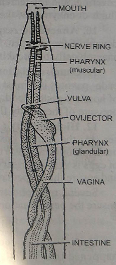
Adult worm is filiform. Body is cylindrical with both ends terminating bluntly. The body is creamish white with smooth and semitransparent body covering. Locomotory organs are absent. Sexes are separate and exhibit sexual dimorphism. Female is 65-100 mm long and 0.25 mm broad. The male measures 40 mm in length and 0.1 mm in diameter. The posterior end of female is straight and blunt, while the posterior end of male is sharply curved and contains genital papillae, two unequal copulatory spicules and caudal alae. The genital pore in female is located in the pharyngeal region. The male genital pores lie at the posterior end of body on the genital papilla. The esophageal bulb is absent.
Life Cycle
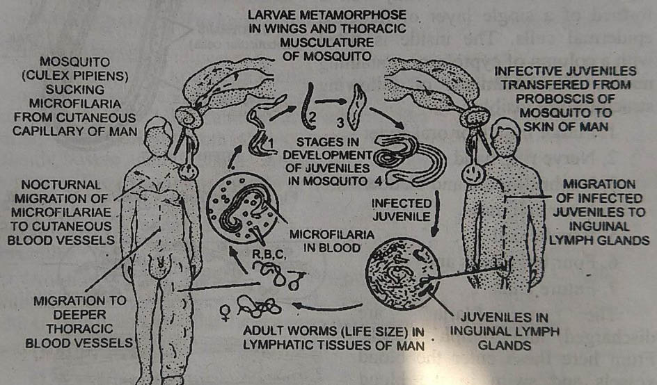
Filaria is digenetic. Its life cycle is completed in two hosts : the primary or final host is man, while the intermediate host is a blood sucking insect the mosquito (Culex or Aedes).
Life cycle in man: Copulation takes place when both sexually mature male and female worms are present in the same lymphatic gland. Filaria is viviparous or ovoviviparous and releases numerous juveniles. These are called microfilaria.
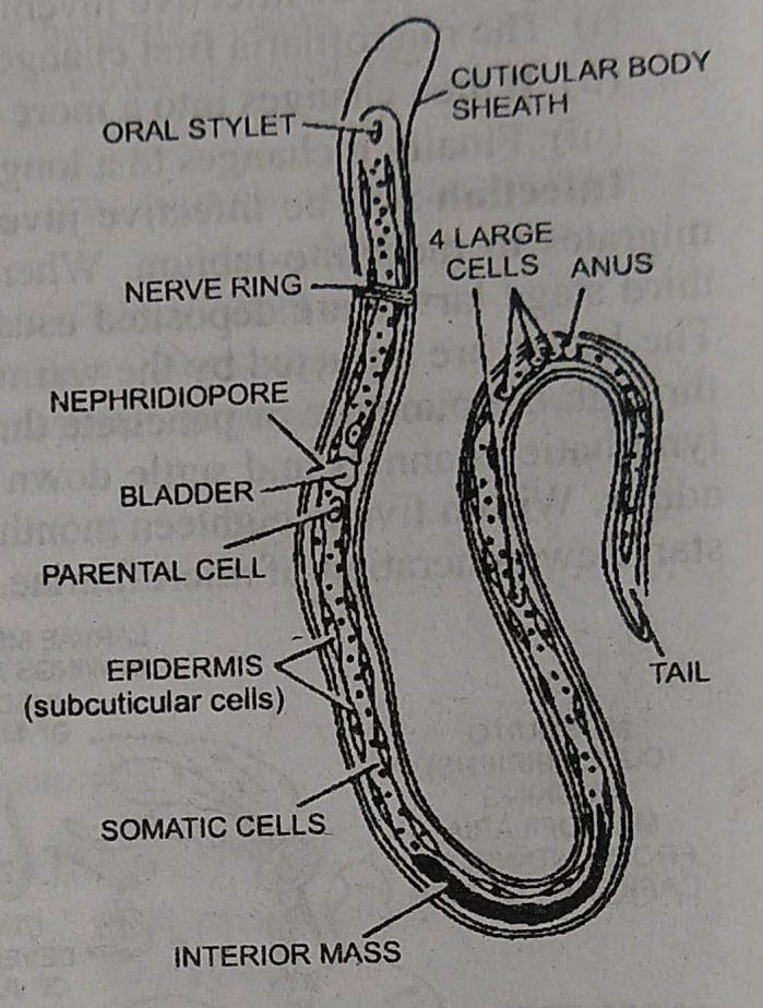
Microfilaria is microscopic about 0.2 to 0.3 mm long. Each is surrounded by a delicate cuticular sheath. Microfilarias contain rudiments of adult organs or structures. The body wall is formed of a single layer of flattened epidermal cells. The inside is filled with a column of cytoplasm containing nuclei. The rudiments of following structures are visible :
1. Future mouth or oral stylet
2. Nerve ring band
3. Nephridiopore and bladder
4. Inner cell mass.
5. Renette cell
6. Four large cells and
7. Future anus
The microfilarias are discharged into lymph vessels. From here these enter the blood vessels and swim in the blood showing active movements. These migrate to deeper blood vessels of thorax. Here these do not develop further until sucked by the intermediate host. If not sucked by mosquitoes, the microfilaria perishes within 70 days.
Life cycle in mosquito: When in blood, microfilaria show diurnal rhythm. During day time, these reside in the deep seated large blood vessels but migrate into superficial peripheral blood vessels in the night. Micro-filaria are sucked by the female mosquito Culex or Aedes.
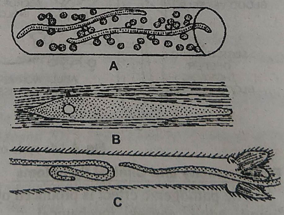
Inside the stomach of mosquito, the sheath is lost and microfilaria penetrates the stomach wall and migrates to the thoracic muscles or muscles of wings. Here microfilaria undergoes two moults in about 10 days to reach the third stage larvae or infective juvenile stage.
(i) The microfilaria first change to a plump sausage-shaped organism.
(ii) It then changes into a more elongated form.
(iii) Finally it changes to a long slender juvenile form.
Infection: The infective juvenile form is about 1.5 um long. It now migrates to mosquito-labium. When the infected mosquito bites a man the third stage larvae are deposited usually in pairs on the skin near the wound. The larvae are attracted by the warmth of skin and they enter the body either through the puncture or penetrate through the skin. From here they reach the lymphatic channels and settle down at some spot to metamorphose into the adults. Within five to eighteen months the adults become sexually mature and start new generation of microfilaria.
Pathogenicity and Clinical Symptoms
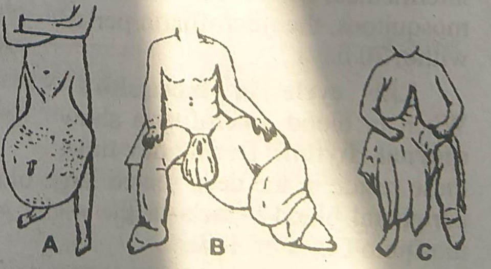
The pathogenic effect is produced by the adult worm living or dead. The living adult causes mechanical irritation and deposits metabolites. As a result the lymph vessels get obstructed. The dead worms also block the lymph vessels. All these initiate proliferation of endothelial cells of lymph vessels leading to inflammatory thickening of the wall of lymphatic vessels. As a result, periodic attacks of fever occur and the tissues surrounding the lymph nodes and other organs of reticuloendothelial system such as liver, spleen, scrotum, vulva, legs and groins become greatly enlarged producing tumour-like solidity. This condition is known as elephantiasis.
Treatment
The drugs used in elephantiasis are divided into three categories depending upon their effect :
(i) On adult worms: Mel W. an or arsenical preparation.
(ii) On microfilaria : Diethyl carbamizine (Hetrazan).
(iii) On infective larvae and immature adult: Paramelamminyl phenylstibonate (MSB).
Prophylaxis
It includes:
1. Destruction of mosquitoes.
2. Protection against mosquito bites.
3. Treatment of carries by using hetrazan.
BSc 1st Year Lower Non-chordates Helminths Sample Model Practice Question Answer Papers