Q.4. Draw a neat and labelled diagram of the longitudinal section of Sycon and describe the different types of cells met in this animal.
Or
Describe the structure and functions of different types of cells in Sycon.
Ans.4. Longitudinal Section of Sycon
Sycon represents cellular grade of organisation. It has two cellular layers, the pinacoderm and choanoderm, with an intermediate mesenchyme.
The pinacoderm controls the inter- relation between the mesenchyme and the external medium, and choanoderm mainly controls the nutrition of the animal.
1. Pinacoderm
It consists of following types :
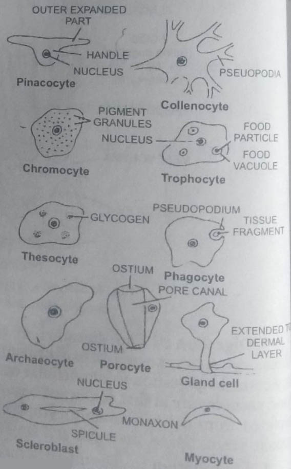
1. Pinacocytes : The pinacocytes are flat and polygonal and scale-like epithelial cells which are cemented together by their thin continuous margins.
Functions: Pinacocytes carry following functions:
(a) These form the external covering of the body except ostia and osculum. These comprise the exopinacoderm or dermal epithelium.
(b) Those lining the incurrent canals and spongocoel form the endopinacoderm.
(c) Since porocytes are highly contractile, they can greatly reduce or increase the surface area of the sponge body.
(d) These engulf the food particles in the incurrent canals.
2. Porocytes : These are tubular cells perforated along their long axis by a central canal, the prosopyle. These are found at frequent intervals among the pinacocytes in the incurrent canals. These are highly contractile and bring the closure of the porocytes. The nucleus lies in the peripheral cytoplasm.
Functions: Porocytes carry following functions :
(a) Form a passage for water current. Water enters the radial canals through prosopyles from the incurrent canals.
(b) By their contraction porocytes regulate the size of the prosopyles.
2. Choanoderm or Endoderm Layer
It constitutes the gastral epithelium and is formed of collar cells or choanocytes.
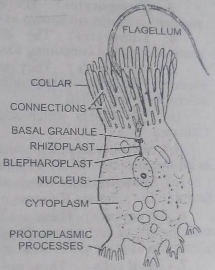
Choanocytes: These are flagellated cells which are oval or rounded in shape. These are loosely arranged upon the mesenchyme. Each choanocyte contains a single nucleus, one or two contractile vacuoles, food vacuoles, reserve food, a bhepharoplast, rhizoplast and a single basal granule or kinetoplast. From the kinetoplast arises a long, whip-like flagellum. The flagellum is surrounded at its base by a thin cytoplasmic collar. It is soft at its tip and somewhat stiff towards its base.
Functions of choanocytes: Choanocytes carry the following functions:
(a) The flagella of choanocytes beat incessantly and make the water current to flow in the body of sponge.
(b) Beating of flagella in a spiral manner draws food particles towards the collar. These adhere to the surface and are captured by the choanocytes to be transferred to the amoebocytes.
(c) The choanocytes also play a role in reproduction by producing sperms.
3. Mesenchyme or Mesohyal
It is a gelantinous matrix lying between the pinacoderm and choanoderm. In the matrix of mesenchyme are found several types of amoebocyte cells. These are modified archaeocytes that migrate from outer layer and carry on a variety of functions. Following types of amoebocytes are found in the body of sponge :
1. Archaeocytes : The archaeocytes are undifferentiated embryonic cells, which play an important role in regenerative processes, and also give rise to sex-cells. These are generalised amoebocytes with blunt pseudopodia, large conspicuous nucleus and often with cytoplasmic inclusions. The archaeocytes participate in the transportation of food and wastes and produce other types amoebocytes and sex-cells.
2. Collenocytes : These are small amoeboid cells with long, branching and anastomosing pseudopodia that form syncytial network. These represent the connective tissue cells.
3. Chromocytes : The amoebocytes with lobose pseudopodia and pigment granules are known as chromocytes.
4. Thesocytes: The thesocytes are amoebocytes having blunt pseudopodia and containing food reserves.
5. Scleroblasts : They are concerned with the secretion of skeletal elements, the spicules. Depending upon the nature of substances secreted, the scleroblasts are called :
(a) Calcoblasts which secrete calcareous spicules and are found in Calcarea.
(b) Silicoblasts that manufacture spicules formed of silica and are found in all the members of class Hexactinellida and some of Demospongia.
(c) Spongioblasts which secrete skeleton of spongin fibres. These are found in the subclass Keratosa of class Demospongia.
6. Myocytes : These are fusiform contractile cells resembling smooth muscle cells of vertebrates. These are arranged in a circular fashion around the osculum and other pores so as to form a sphincter to regulate their opening.
7. Gland cells: These are found attached to the body surface by means of long strands. These secrete lime.
BSc 1st Year Lower Non-chordates Sycon Sample Model Practice Question Answer Papers