Q. 2. Describe life history of Taenia.
Describe life-history and economic importance of Taenia.
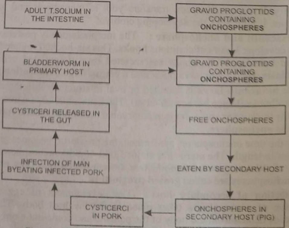
The life-history of Taenia is complicated and digenetic, being completed in two hosts. The primary host is man and the secondary host is pig.
Fertilisation : Self-fertilisation occurs as a rule. The cirrus of the segment is inserted into the vagina of the same segment. The sperms received are stored in the receptaculum seminis. The eggs are fertilised in the oviduct.
Copulation occurs only if gonopores of two mature proglottids come in contact due to folding of strobila. During copulation cirrus is inserted into the vagina and sperm are transferred.
Capsule Formation
In ootype each fertilised egg or zygote becomes associated with a large yolk cell or vitelline cell secreted by the vitelline gland. The zygote and yolk cells are enclosed in a thin shell or chorionic membrane. It is formed by the material exuded by the yolk cell.
The capsulated eggs are collected in the uterus, where these undergo further development. Passage of capsules into uterus is lubricated by secretion of Mehlis glands. To accommodate developing embryos, the uterus becomes highly branched.
Eggs : The eggs are very small measuring about 40 microns in diameter. These contain a large amount of yolk and each is surrounded by an egg-shell or egg-capsule.
Development
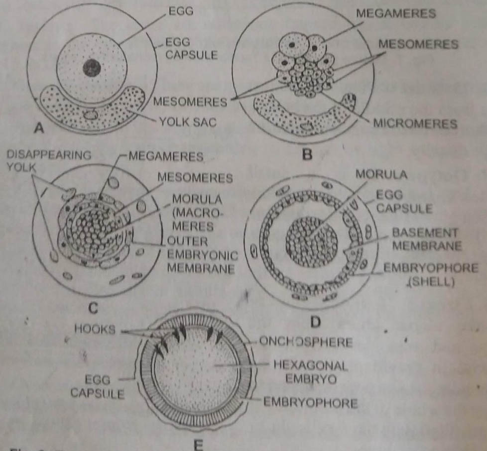
Cleavage: The zygote starts dividing while still inside the uterus. The first cleavage is holoblastic but unequal so that a large megamere and a small embryonic cell are formed.
Morula : Megamere divides several times forming a number of megameres. The embryonic cell divides repeatedly and produces two types of embryonic cells: larger mesomeres and smaller micromeres. (i) Micromeres form a rounded mass of cells and called morula. (ii) Mesomeres form a layer outside the morula. It develops inner envelope and finally forms a thick, hard radially striated shell, and is called embryophore or inner embryonic membrane. (iii) Megameres form outer envelope or outer embryonic membrane.
The yolk or vitelline cell transfers yolk to megameres and gradually disappears. It nourishes the developing embryo.
Hexacanth and onchosphere: The morphologically posterior end of morula develops 3 pairs of chitinous hooks. This six-hooked rounded embryo is called hexacanth. Its hooks are secreted by differentiated cells, called onchoblasts. Hexacanth larva possesses a pair of large penetration glands.
The hexacanth larva is rounded or oval in structure with three pairs of chitinous hooks (hence hexacanth larva). The hexacanth larva when enclosed inside the thick inner and outer thin embryonic membranes (embryophores) is known as onchosphere.
By the time onchospheres are formed, the proglottid becomes gravid and increases in length. The uterus has about 3,000-40,000 onchospheres. Except uterus, all other organs of reproductive system degenerate. The proglottids with onchospheres are called gravid proglottids.
Infection of secondary host : The development of egg upto the formation of onchospheres takes place inside the host body. The gravid proglottids detach from the body of the parasite and come out along with the host faeces. These infect the secondary host when pig feeds upon the contaminated faeces. Dog, camels or monkeys are also infected sometimes and even auto-infection of man is also possible.
Cysticerus or hydatid larva or bladderworm stage: The numerous hexacanths are set free in the stomach, where the embryonic membranes on onchospheres are dissolved.
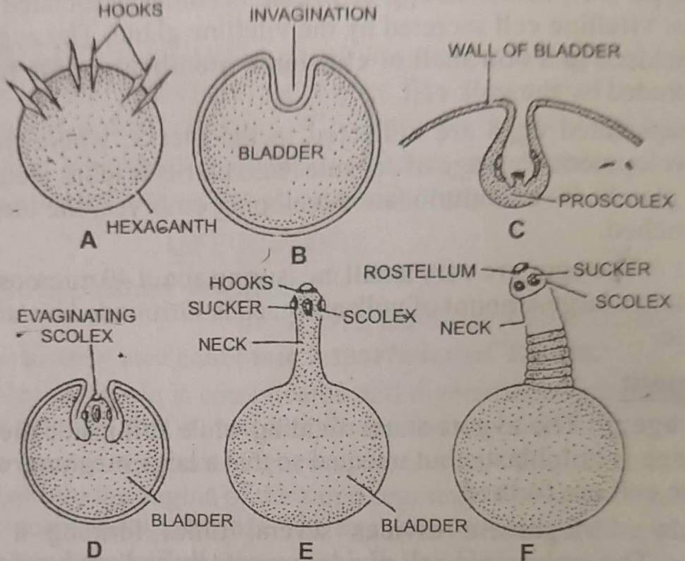
These bore through the intestinal wall with the help of hooks and enter the blood stream of lymph vessels. Travelling through the heart, these enter the muscles of various parts in the body. The usual site where the hexacanths get encysted are the voluntary muscles of tongue, heart, liver and shoulder.
In the muscles, the hexacanth loses hooks, increases in size and develops a central fluid-filled space. As such a single layered sac-like structure, the bladder, is formed. It is formed by an outer cuticle and an inner mesenchyme. As the bladder increases in size an invigination occurs on one side. The inpushing differentiates as a hollow knob with which the development of suckers and hook on its inner side is converted into the proscolex.
Finally, the proscolex is everted and the hooks and suckers are exposed to the exterior. It is now known as bladderworm or cysticercus larva. It is in the form of an oval vesicle with a small scolex similar to that of an adult having four suckers. Many cysticerci can be seen as white dots in the flesh of pig which is now known as measly pork.
Infection of primary host : Further devlopment of the bladderworm is not possible inside the pig and can take place only if taken by the definitive host.
Infection of man occurs when inadequately cooked pork infected with bladderworms is eaten. The cysticerci become active in the intestine. The scolex takes a firm hold of intestinal wall of the host. The bladder is thrown off and the neck starts budding off proglottids and an adult tapeworm is formed. It becomes sexually mature in about 2 to 3 months.
BSc 1st Year Lower Non-chordates Taenia Solium Sample Model Practice Question Answer Papers