BSc 2nd Year Zoology Development of Tongue Worm Notes Study Material
BSc 2nd Year Zoology Development of Tongue Worm Notes Study Material: BSc is a three-year program in most universities. Some of the universities also offer BSc Honours. After getting enrolled for BSc, there are certain things you require the most to get better grades/marks in BSc. Out of those, there are BSc Study Material, BSc Sample Model Practice Mock Question Answer Papers along with BSc Previous Year Papers. At gurujistudy.com you can easily get all these study materials and notes for free. Here in this post, we are happy to provide you with BSc 2nd Year Zoology Development of Tongue Worm Notes Study Material.

BSc 2nd Year Zoology Development of Tongue Worm Notes Study Material
Introduction
Asexual reproduction is a rare phenomenon in hemichordates but Gilchrist (1923) reported asexual reproduction in Balanoglossus capensis. In summer months isolated parts of the tail of the juvenile phase develop completely into adults in subsequent winters. During sexual reproduction zygote of Balanoglossus develops either directly or indirectly depending on its size. The small fertilized eggs develop indirectly but large fertilized eggs develop directly. The indirect development is through a larval form called tornaria larva. In both cases, the early phases of development are the same. (BSc 2nd Year Zoology Development of Tongue Worm Notes Study Material)
Fertilization
During the breeding season (May to June), mature sperms and ova escape into the surrounding sea water where fertilization takes place. Thus, fertilization is external. (BSc 2nd Year Zoology Development of Tongue Worm Notes Study Material)
Indirect Development
The indirect type of development is studied in Balanoglossus clavigerus by workers like Heider (1909), Stiansy (1914), and Payne (1937).
Embryonic or pre-larval development
The pre-larval development is similar to that of Branchiostoma. The zygote, produced as a result of fertilization, undergoes cleavage which is holoblastic, almost equal, and of radial type. It results in a sphere of blastomeres, the morula. The first cleavage is vertical. The second is also vertical but at a right angle to the former. The third cleavage is horizontal and results in 8 blastomeres of unequal sizes, the upper ones are smaller micromeres and the lower ones are larger macromeres.
The morula undergoes reorganization of its blastomeres and takes the form of a single-layered, hollow, and spherical embryo, the blastula or coeloblastula. Its central fluid-filled cavity is called the blastocoel. Blastula results in about 6-15 hours after fertilization. Within 12 to 24 hours, an invagination starts in the blastula which deepens to form the archenteron that opens outside through a blastopore. Soon the blastopore closes and the embryo, now called gastrula, lengthens along the anteroposterior axis. Now the tip of the archenteron is pinched off as a coelomic vesicle called the protocoel. Thus origin of coelom is enterocoelic.
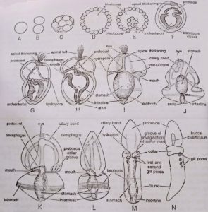
Balanoglossus Stages of DevelopmentThe remaining portion marks the future gut. The protocoel becomes triangular in shape. Its one end gets attached to the underside of the apical thickening and another end opens outside through an aperture, the hydropore, towards the dorsal side of the embryo. The protocoel and hydro pores are the future proboscis coelom and probosci’s pores, respectively. The collar and trunk coelom arises as solid invaginations of the hindgut, independent of the formation of protocoel.
Larval development
With the formation of the protocoel, the inner ends of the early gut move towards the ventral surface and open to the outside through a mouth. The gut is now regional into the esophagus, stomach, and intestine. The intestine opens outside through an anus, formed at the side of the closed blastopore. By this time the embryo becomes uniformly ciliated and escapes from the egg membrane on the 7th day to lead a free-swimming larval life. It is called tornaria. (BSc 2nd Year Zoology Development of Tongue Worm Notes Study Material)
Tornaria larva
The Tornaria larva was first described by J. Muller in 1850 who suspected it to be the larva of some starfish. Later on, it was known to belong to Balanoglossus clavigerus. It is so-called because of its habit of rotating in circles. It is clear, and glossy in appearance with an oval body ranging up to 3 mm in size. It has a ventral mouth near the equatorial plane of the body, a posterior terminal anus, and a gut differentiated into an esophagus, stomach, and intestine. The cilia form two bands on the body surface.
The anterior ciliary band or circumoral band takes up a winding course over the preoral surface and forms a postoral loop, its cilia are short and serve to collect food. The posterior ciliated band or telotroch occurs as a ring in front of the anus, its cilia are long and serve as locomotor organs. At the anterior end is an apical plate of thickened epidermal cells, which bears a pair of eye spots or ocelli and a tuft of sensory cilia called apical tuft or ciliary organ eyes are cup-shaped, cavity of the cup is filled with clear material constituting the lens.
Cells of the cup are provided with pigment granules. The protocoel (proboscis coelom) in the form of a thin-walled sac, is present and opens to the exterior through a hydropore (proboscis pore). To the right of the hydropore lies a pulsating heart vesicle. The collar and trunk coeloms appear in the older larva. (BSc 2nd Year Zoology Development of Tongue Worm Notes Study Material)
Metamorphosis
The larva swims freely, leads a planktonic life, feeding on minute organisms, and metamorphoses into an adult worm. During metamorphosis, the size is reduced probably due to the loss of water. Transparency, ciliary bands, sensory cilia, and eye spots are lost. The body becomes differentiated into proboscis, collar, and trunk by the appearance of two constrictions, and the trunk region is elongated.
The hydropore persists as proboscis pore, and the buccal diverticulum and gill-slits appear as outgrowths of the alimentary canal. Reproductive organs mark their presence, probably they develop from mesoderm. The tongue bar grows. An adhesive post-anal tail is formed which helps in anchoring the young worms and later resorbed. The animal now sinks to the bottom to lead a benthonic life as an adult. (BSc 2nd Year Zoology Development of Tongue Worm Notes Study Material)
Direct Development
Direct development is found mostly in the genus Saccoglossus. It is found in the large eggs provided with a good amount of yolk. In this case, a tornaria larva is not formed. The Blastopore of the gastrula narrows down and finally closes.
Cilia develop on the general surface of the gastrula stage of the embryo. Later embryo becomes free and swims in water for about 24-26 hrs. The embryo elongates and becomes more worm-like. A transverse groove appears and the mouth develops by invagination on the position of the groove. (BSc 2nd Year Zoology Development of Tongue Worm Notes Study Material)
The anus develops at the position earlier occupied by blastopore. Before the formation of the mouth, a diverticulum is separated in the anterior region from the archenteron. The cavity of this forms the proboscis coelom. Paired collar trunk coelom is formed as an outgrowth from the posterior wall of the proboscis cavity directly from the walls of the archenteron. At this stage, structures elongate and get differentiated into proboscis, collar, trunk, etc. (BSc 2nd Year Zoology Development of Tongue Worm Notes Study Material)
BSc 2nd Year Zoology Development of Tongue Worm Notes Study Material
BSc 2nd Year Sample Model Practice Mock Test Question Answer Papers