Microbiology Viral Diseases of Man Notes Study Material
Microbiology Viral Diseases of Man Notes Study Material: We provide BSc 2nd Year Microbiology Notes Study material, question answers, sample papers, mock test papers, and pdf. At gurujistudy.com you can easily get all these study materials and notes for free. Here in this post, we are happy to provide you with BSc 2nd Year Microbiology Viral Diseases of Man Notes Study Material.

Microbiology Viral Diseases of Man Notes Study Material
Viruses pathogenic to humans are artificially classified into four mainly on the basis of organs on or in which typical symptoms a produced. These groups are as follows:
1. Pneumotropic viral diseases, involving the respiratory tracts, such as influenza and common colds.
2. Dermotropic viral diseases, involving mainly skins, such as measles chicken pox, smallpox, etc.
3. Viscerotropic viral diseases, involving mainly internal organs, such as yellow fever, dengue fever, hepatitis, etc.
4. Neurotropic viral diseases, involving mainly the central nervous system, such as rabies, polio, encephalitis, etc.
In each of the above four groups, other organs may also be involved. We shall explore some of the common diseases in each group.
[I] Influenza
This is an acute disease of the upper respiratory tract. The virus is a helical virion containing single-stranded RNA and is enveloped. The envelope contains a series of projections or spikes as shown in Figure.
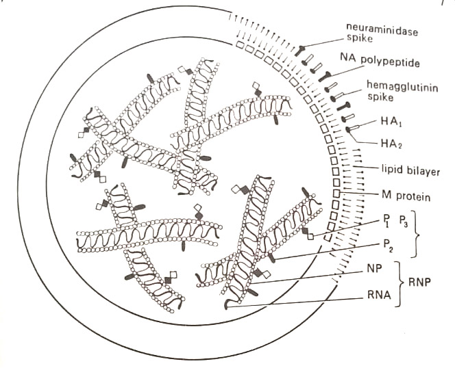
These spikes contain the enzymes hemagglutinin which allows the virion to bind the red blood cells and neuraminidase which dissolves the cell membrane during replication. Both enzymes are said to be antigens because they induce the body to form antibodies that neutralize the virion. The hemagglutinin spike is composed of three sets of HA and HA, polypeptides. The neuraminidase spike consists of four NA polypeptides. The nucleocapsid is segmented and gives the appearance of a double helix due to the association of internal proteins (NP5, P1, P2, and P3).
There are three distinct types of influenza virus: Types A and B cause worldwide epidemics called pandemics, and Type C, which occurs sporadically. Each type is known for its antigenic variation, a process in which changes occur in the protein structure of the capsid as well as in the hemagglutinin and neuraminidase. This results in a myriad of variants that are unrecognized by the antibodies produced in previous infections. One, therefore, suffers innumerable cases of influenza.
Also, this phenomenon prevents the development of an effective vaccine. The nomenclature for influenza viruses is derived from the changing antigenic pattern. Virologists name the viruses according to their type, the animal of origin other than humans, the location of the first isolation, the strain number, and the year of isolation. It is thus common to see such names as A/Victoria/3/75 or A/Swine/ Wisconsin/15/30. (Microbiology Viral Diseases of Man Notes Study Material)
The infection causes sudden chills, fatigue, headache, and general aches, and pains mainly in the back and legs. The fever may rise to 103°F – 104°F over a 24 hr period, with a severe cough. The nose obstructs, the throat becomes dry, and the chest is tight. Transmission occurs usually by bits of respiratory mucus and salvia called droplets. There may be secondary bacterial infections – Streptococcus and Staphylococcus invading and damaging the epithelium. The disease is diagnosed by testing patients’ serum for neutralizing antibodies.
If the titer or concentration of antibodies increases during the course of infection, the disease is confirmed. Hemagglutination tests are also made.
Influenza A is sometimes treated with amantadine, a synthetic drug that may block the viral penetration to cells. In an epidemic state, influenza may result in Guillain-Barre syndrome (GBS) causing nerve damage and polio-like paralysis, and Reye’s syndrome involving the liver and brain, especially in young children. (Microbiology Viral Diseases of Man Notes Study Material)
[II] Adenovirus infections
Adenoviruses are a collection of at least 35 types of icosahedral virions having double-stranded DNA. The virion is 60-90 nm in diameter, having a dense central core and an outer coat, the capsid. The capsid is composed of 252 capsomeres – 240 hexons (making the faces and edges of equilateral triangles) and 12 pentons (making the vertices). In capsid, there are four additional minor proteins (IIIa, VI, VIII, IX) associated with the hexons or pentons in stoichiometric amounts. The core proteins include proteins V and VII associated with the viral DNA.
The viruses owe their name to the adenoid cells from which they were first isolated in 1953. They multiply in the nucleus of infected cells causing visible nuclear granules or inclusions, which are made up of numerous virions arranged in a crystalline pattern. Adenoviruses cause diseases of the respiratory tract, eyes, and meninges. (Microbiology Viral Diseases of Man Notes Study Material)
Respiratory infections are transmitted by droplets. Like influenza, they also cause mild hoarseness, cough, and loss of appetite called anorexia. The terms common cold and croup are often used for infections. Type 8 adenovirus is the chief cause of kerato-conjunctivitis-eye inflammation, tearing, sensitivity to light, and swelling. Type 7 causes meninges – in the spinal cord coverings.
[III] Rhinovirus infections
They are a large group of more than 100 small RNA viruses with icosahedral symmetry. Rhinos means nose. The virion consists of a single molecule of RNA and a capsid containing 60 copies of four different polypeptide chains. They are involved in respiratory infections. They multiply under the respiratory tract causing injury to mucosal cells. There is a headache. sneezing, stuffy nose, and sore throat, a condition called acute coryza, but more commonly as a head cold. There is burning of the eye and nasal membranes with little fever.
Of the many types, only 15-20 cause most infections in humans. Vitamin C and chicken soup are useful. Interferons are being developed. For instance, zinc ions are found to mask viral proteins and inhibit multiplication.
They are generally transmitted through droplets. However, it may also be due to contact and through fomites.
[IV] Chickenpox (Varicella)
This is among the most communicable diseases caused by the varicella-zoster virus. The virus belongs to the herpesvirus group. It is a double-stranded virion with icosahedral symmetry and 162 capsomeres. In adults, the same virus causes herpes zoster (shingles).
The virus is transmitted chiefly by droplets and skin contact. There is the beginning of fever, anorexia, and headache when the virus multiplies in the respiratory tract. When it passes to the bloodstream and localizes in the peripheral nerves and skin, it multiplies rapidly in cutaneous tissues. This results in the development of fluid-filled, tear-drop-shaped skin lesions called vesicles. The vesicle contains a large amount of virus-laden, highly infectious fluid. Later, after 3-4 days, they break open, forming crusts that become dry.
The lesions are larger than those of smallpox. The disease becomes serious if the lungs ( in which pneumonia may develop) or brain (encephalitis) are involved. Secondary infections of the skin by bacteria may also occur. Adenine arabinoside-A (Ara-A), a nucleic acid substitute has shown good results in the treatment. In Japan, a vaccine is also developed.
Herpes zoster (shingles) in adults is a painful disease in which the virus multiplies in the ganglia along the spinal cord and then passes down the nerves of the trunk to the skin. There are superficial tingling and burning sensations in the skin with blotchy red patches.
[V] Herpes simplex
Herpes simplex is a collection of diseases. It may occur as cold sores in the mouth, eczema of the skin, encephalitis in newborns, or urinogenital infections in adults. The virus is also linked with human tumors.
There are two major types of herpes simplex virus: Type 1, designated as HSV-1, infecting usually areas above the waist, whereas Type-2, HSV-2, is often found below the waist. (Microbiology Viral Diseases of Man Notes Study Material)
Both are large DNA viruses with icosahedral symmetry and envelopes with spikes The particles have a diameter from 180 nm to 200 nm. The DNA-protein core consists of double-stranded DNA wrapped around an associated protein, as one the spindle of a spool. The envelope contains a number of glycoproteins, the capsid is composed of 162 elongated hexagonal prisms, the capsomeres, and at least four unique proteins; and the so-called integument consists of about eight distinct polypeptides.
The viruses multiply rapidly in the nucleus of infected cells forming inclusions called Lipshutz bodies, which aid in viral diagnosis. The virus remains for a long period of time in the body causing recurrent infections in spite of the presence of antibodies. The Greek word herpes means to creep, indicating the spreading nature of the disease. (Microbiology Viral Diseases of Man Notes Study Material)
In adults, herpes simplex infections appear as closely grouped, thin-walled vesicles, or cold sores which occur repeatedly in the same general area of skin or mouth. In children, the disease manifests itself as cold sores or as a more serious condition, gingivostomatitis. Type 1 herpes simplex virus also induces a type of nonepidemic encephalitis in children. Viral multiplication in the brain tissue leads to lesion formation. Transmission occurs by several means including contact with saliva or contaminated utensils.
Adults commonly suffer a genital form of herpes simplex caused by HSV-2, and lesions develop on the external genital organs of the male, and internally along the vaginal wall and cervix in females. In this case, transmission is by sexual contact. The child borne by an infected female may develop herpes encephalitis as the virus is transmitted from the vaginal fluids.
In recent years, the acronym TORCH has been coined for diseases with congenital significance – T for toxoplasmosis; R for rubella; C for cytomegalovirus infection, and H for herpes simplex. The O represents other diseases. HSV-2 is found to be associated with cervical tumors.
A nucleic acid analog, iododeoxyuridine (IDU), replaces thymine in the DNA of the virus and produces a nonsense viral genome. This is applied topically to reduce side effects and used for herpes simplex infections of the skin as eye lesions and cold sores. Adenine arabinoside (Area-A) also known as Vidavabine (Viva-A) is also applied as a topical ointment. Acyclovir (Zovirax) was licensed in 1982 against all herpes viruses, especially for genital simplex. The herpes virus is one of the most common viruses in the environment.
[VI] Measles (Rubeola)
This is a highly communicable respiratory disease in children, whose symptoms develop on the skin. Rubeola is derived from the Latin rube, meaning red- indicating the red rash of the disease. In the beginning, there is fever, coughing, and sneezing, the most communicable phase of the disease. As the virus localizes in subcutaneous tissues, the skin breaks out in a blotchy rash. The rash begins behind the ears at the hairline, then covers the face and trunk. (Microbiology Viral Diseases of Man Notes Study Material)
The first evidence of disease appears along the gums and on the wall of the pharynx. There develop red patches with central white lesions. These occur concurrently with the fever and are known as Koplik spots, after the American physician, Henry Koplik who first described them. This is an important symptom. (Microbiology Viral Diseases of Man Notes Study Material)
The virus is a helical RNA virion, closely related to the mumps and RS viruses. The average size is 125-250 nm (range 100-800 nm), nucleocapsid diam. 18nm, single-stranded RNA of mol. wt. of 5-6 x 10⁶ daltons. It contains envelope spikes with hemagglutinin, a factor used in identification by the hemagglutination-inhibition (HI) test.
There are no neuraminidase spikes on the envelope. Measles may lead to many complications. Due to secondary infection of the damaged epithelium of the respiratory tract, bacterial pneumonia may also develop. There is also evidence that the measle virus may be related to multiple sclerosis, diabetes, and encephalitis. (Microbiology Viral Diseases of Man Notes Study Material)
The earliest vaccine for measles was developed in 1954 from chemically inactivated viruses by John Enders and Thomas Peebles. It was, however, replaced in the 1960s by another vaccine containing attenuated viruses. At present, the children are vaccinated after 15 months of age.
[VII] Mumps
This is a disease of children’s salivary glands, especially the parotid glands – thus the technical name, epidemic parotitis. The disease is generally transmitted by droplets, contact, and fomites. The virus is present in urine, blood, and cerebrospinal fluid. The disease may begin on one side of the mouth but both glands are affected in most cases. This virus occurs only in humans. It is a single-stranded – containing helical virion, and closely related to measles and RS viruses respects the virions are similar to the measles virus except that there are neuraminidase spikes on the envelope. (Microbiology Viral Diseases of Man Notes Study Material)
There is hemagglutinin-containing spikes on the envelope. It is usually a benign disease without any complications in children. There develop antibodies. In adults, mumps represents a threat to reproductive organs, and some males may develop orchitis, i.e. swelling of the testicle to three or four times normal size. Sperm count may be reduced. Though rare, in females, oophoritis (lower back pain and enlargement of ovaries) may develop. (Microbiology Viral Diseases of Man Notes Study Material)
The mumps vaccine was developed in 1967 from live viruses of the Jeryl Lynn strain grown in duck embryo tissues. The virus may multiply in other tissues, in beta cells of the pancreas, causing pancreatitis, or in brain tissues causing mumps encephalitis.
[VIII] Small pox (Variola)
It has a complex architecture without obvious symmetry. The virus is a brick-shaped DNA particle, largest in size – 270 nm. The nucleocapsid is surrounded by a swirling series of fibers and has no envelope. The particles have rounded corners and a central dense region with crescentic dense areas on each side. The surface layer is made of threadlike, double-ridged, beaded structures.
In section, there appears a central nucleoid with a dumbbell-shaped dense core composed of regularly arranged, DNase-sensitive, electron-dense threadlike structures. The nucleoid is surrounded by lipoprotein membranes. Between the nucleoid and the outer viral coat is an ellipsoidal body that causes a prominent central bulging of the virion. The article is composed of about 3.2% DNA associated with spermidine, 91.6% protein, 5% lipid, and 0.2% non-DNA carbohydrate (present in the membrane glycoproteins).
The early symptoms are chills, fever, and general prostration. With the fall in temperature, pink-red spots called macules to appear, first on the scalp and forehead, then on the neck and extremities, and finally on the trunk. The spots now turn to pink pimples or papules, and later fluid-filled vesicles. The technical name, variola is derived from the Latin varus meaning pimple. In the final stage, the vesicles advance to pustules, covering the body surface. When they break open, the pus comes out.
They are deep in the skin rather than on the surface as in chicken pox. The pustules may form a soft crust, which falls off leaving a pitted scar, or pock. The name smallpox was used to distinguish the pock from the larger lesions of syphilis or varicella (chicken pox).
Transmission is by contact with the skin or any of the body fluids including blood, urine, or droplets. In 1798, an English physician, Edward Jenner observed that milkmaids and others working with dairy cattle contracted a closely related but mild form of pox called cowpox, or vaccinia from the Latin Vacca means cow. (Microbiology Viral Diseases of Man Notes Study Material)
Apparently, those who had recovered from cowpox did not contract smallpox. Jenner, therefore, substituted material from a cowpox lesion for the smallpox material and established the process of vaccination. His method became so famous that Napoleon ordered his army to be vaccinated in 1806.
The introduction of the cowpox virus to the bloodstream induces the body to manufacture antibodies that neutralize both the cowpox and smallpox viruses as they are partially similar. The World Health Organisation (WHO) received 1967 funds for an attempt at the global eradication of smallpox. By a process of Surveillance containment, pox patients were isolated, and every known case was vaccinated. The last epidemic occurred in 1974 in Bangladesh, and in 1976 in Ethiopia. By October 1977, the WHO claimed that the last case had been isolated.
On October 26, 1979, the announcement was made that smallpox had been eliminated from the earth, the first such claim made for any disease. The virus is said to exist only in four research laboratories in the U.S.A., Russia, the U.K., and Japan, permitted by the WHO.
[IX] Dengue fever
There are four distinct types of virions of dengue fever. This virus is an RNA virion of apparently icosahedral symmetry and with an envelope. The virus is transmitted when a diseased mosquito injects its saliva into the wound during a blood meal. Only the female bites for her need for human blood for egg production. The mosquito becomes infectious nine to twelve days after it has consumed blood from the dengue fever victim, and it will soon die regardless of whether it transmits the virus. (Viral Diseases of Man Notes Study Material)
Dengue fever is accompanied by severe fever and prostration. There is severe pain in limbs and muscles and one feels as if bones are breaking-breakbone fever. Immunity to a particular strain of virus is lifelong. Due to infection, the person may develop dengue hemorrhagic fever (DHF). There is facial rash, severe vomiting, and circulatory failure due to shock. This disease has been traditionally found in areas of the southeast Pacific and in Southeast Asia. Later it also broke into central America and southeast Mexico.
[X] Rabies
Rabies has the highest mortality rate of any human disease once the symptoms have fully materialized. It occurs in most animals in nature from dogs and cats to horses and rats, to skunks and bats. It is equally fatal to animals and humans.
The virion is RNA containing enveloped particles, rounded on one end and flattened on the other to give the appearance of a bullet. The virus enters the tissue during a bite, skin wound, or abrasion from an infected animal. Though, a disease associated with saliva, urine, lymph, blood, or milk may be equally infectious. In 1978, a woman died of rabies in Boise, Idaho, due to a cornea transplant from an infected person.
The incubation period for rabies varies according to the number of virions introduced to the wound and the wound’s proximity to the central nervous system. It may vary from six days to one year, and is generally shorter in children than in adults. The virus multiplies in the muscle tissue and then spreads rapidly to the neural pathways. Early signs of rabies are tingling, burning, or coldness at the bite site along with fever and headache. Later, due to increased muscle tone, the patient becomes alert, aggressive, and shows unusual behavior.
Due to the paralytic effect on pharyngeal muscles, difficulty in swallowing is felt. Salivation becomes profuse and saliva drips from the mouth as it cannot be swallowed. This symptom together with brain degeneration increases the person’s reactions to the sight, sound, or thought of water. Traditionally, the disease has been called hydrophobia, meaning “fear of water”. The paralysis spreads and death occurs within a few days of respiratory inhibition. (Microbiology Viral Diseases of Man Notes Study Material)
Dogs are usually infectious for ten days before they show rabies symptoms. Due to this, a dog may be held for observation after a bite. If ten days pass without any event it may be assumed that the dog was not contagious at the time of the bite. If the dog dies immediately after the bite, its brain tissue sample could be inoculated to laboratory mice that should show symptoms within three weeks if the dog was rabid. A person suffering from an animal bite should be precautionarily and treated as if the animal were rabid.
The wound is flushed thoroughly with soap and any antiseptic is applied. Serum containing anti-rabies antibodies should be injected into the base of the wound. Thus interferon is helpful. A tetanus shot may also be given if not given during the last ten years or so.
A combination of vaccine and antibodies are then injected to neutralize the virus before it reaches the central nervous system. Early vaccines contained viruses from chick embryos or rabbit nerve tissue. But these were found to be allergic. Later, a vaccine was produced from viruses cultivated in duck embryo tissue inactivated with propiolactone. Duck embryo vaccine (DEV) was injected into the abdominal fat at a 45° angle every 14 successive days or more if needed. (Microbiology Viral Diseases of Man Notes Study Material)
This virus is absorbed slowly from among the fat cells and exposes the body to the virus for a longer period. The injections were painful due to the sensitivity of the abdominal muscles.
Recently, a new rabies vaccine, Merieux human diploid cell vaccine, is shown to produce a high amount of antibodies after only three injections in the arm. The viruses are cultivated in human embryonic lung cells, and since human tissue is used, allergic responses are minimum. This vaccine, licensed in 1980 in the U.S.A is now used in other countries including India. It is an expensive vaccine.
Rabies in animals occurs in two forms:
1. Furious form. Characterized by violet symptoms as the animal becomes wide-eyed, drools, and attacks anything in sight. It becomes paralyzed, lapses into a coma, and dies. (Microbiology Viral Diseases of Man Notes Study Material)
2. Dumb form. Recognized by deep lethargy and dies suddenly of paralysis.
[XI] Polio
This is one of the smallest known viruses. It has a diameter of about 27-30 nm. It has single-stranded RNA (mol. wt. 2.5 x 10⁶ daltons i.e. 7.7 Kb) with an icosahedral capsid. It multiplies rapidly in nerve cells, producing over 10,000 new particles during a single cycle of about six hours.
The virus enters the body through contaminated food or water. It first multiplies in the tonsils and the lymphoid tissues of the gastrointestinal tract. There may be influenza, nausea, vomiting, and intestinal cramps. The disease may end at this point. However, in some cases, it progresses, and the virus passes to the bloodstream. It then localizes on the gray matter of the spinal cord. The word polio is taken from the Greek polio meaning gray. Meningitis may occur and paralysis experienced in the muscles of the arms, legs, trunk, and other areas controlled by the spinal cord. This condition is often called aseptic meningitis.
The most serious form of polio is the one in which the virus infects the stem of the brain, known as the medulla. This condition is called bulbar polio, due to the bulblike appearance of the medulla. The nerves of the upper body torso are affected. Swallowing becomes difficult and paralysis occurs in the tongue, facial muscles, and neck. (BSc Microbiology Viral Diseases of Man Notes Study Material)
Three types of poliovirus are known to exist. Type I, the Brunhilde strain, causes a major number of epidemics and moderate cases of paralysis. Type II, the Lansing strain, occurs sporadically but induces the greatest percentage of paralytic cases. Type III, the Leon strain, also occurs sporadically but the virus is confined to the intestine, rarely causing paralysis.
Jonas Salk and his group prepared 1940s huge quantities of viruses and inactivated them with formaldehyde. The virus was then injected into the body to produce antibodies. Albert Sabin and his group developed a vaccine containing attenuated viruses prepared by multiple passages through tissue cultures. This vaccine may be taken orally and is in current use. It produces more antibodies, providing a longer exposure to the virus due to the low multiplication rate of viruses in the intestine. Modern vaccines incorporate all three types of polioviruses, said to be trivalent. (BSc Microbiology Viral Diseases of Man Notes Study Material)
AIDS: A Death Warrant
The first of the few cases of AIDS was reported in 1981 by the Centre for Disease Control, Atlanta, USA. Isolated cases of AIDS were identified as early as 1976. Serological data suggest that the AIDS virus was not present in the human population before 1979. There is suspicion that this virus may be a monkey virus which was first transmitted to man in Africa and then to the USA and Europe. Millions of Australia, South Africa, Central Africa, different parts of Europe, the Caribbean, and Japan.
Reports of the incidence of AIDS are increasing in various parts of India also. The disease that was once restricted only to the west, is gradually appearing here also. Medical researchers have been examining blood samples of the suspected individuals.