BSc 2nd Year Microbiology Viruses Notes Study Material
BSc 2nd Year Microbiology Viruses Notes Study Material: BSc is a three-year program in most of universities. Some of the universities also offer BSc Honours. After getting enrolled for BSc, there are certain things you require the most to get better grades/marks in BSc. Out of those, there are BSc Study Material, BSc Sample Model Practice Mock Question Answer Papers along with BSc Previous Year Papers. At gurujistudy.com you can easily get all these study materials and notes for free. Here in this post, we are happy to provide you with BSc 2nd Year Microbiology Viruses Notes Study Material.

BSc 2nd Year Microbiology Viruses Notes Study Material
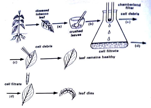
Although viral diseases like smallpox and yellow fever are known since the early days of human history, the nature of their causes was, however, known quite late. Adolf Meyer, a Dutch agricultural chemist in 1886 observed a mottling disease in the leaves of tobacco plants and named it Mosaikkrankhet i.e. mosaic. He could show the infectious nature of the sap of diseased leaves.
He was able to demonstrate that the clear filtrate obtained by passing the juice (obtained by grinding the diseased leaves in water) through double filter paper was infectious (i.e. caused disease when applied to healthy leaves). Since the capacity of causing the disease was lost by heating at 80° C, he concluded that bacteria were the cause of the disease.
Iwanowski, a Russian in 1892 infact was the first to discover a viral disease in plants in the sense that he could demonstrate for the first time that tobacco mosaic could be transmitted to healthy plants through the sap from diseased plants even though the sap had been passed through the filters fine enough to remove all bacteria (Chamberland filter candles).
However, no living thing capable of producing tobacco mosaic grew from the filtered sap of diseased plants on any culture medium. Different steps of his experiment are shown in Figure Beijerinck in 1898 in Holland found that the filterable, invisible, and non-cultivable infectious principle would diffuse through an agar gel, like a fluid. He thought the fluid itself alive and called it contagium vivum fluidum i.e. a living infectious fluid. (BSc 2nd Year Microbiology Viruses Notes Study Material)
Viral diseases of vertebrates were well known by 1892 since Louis Pasteur had been studying canine rabies for some time. He indicated the cause of this disease as a virus. The term virus was then commonly used for a variety of infectious agents, including bacteria.
Loeffler and Frosch in 1898, showed that the agent of foot-and-mouth disease of cattle, like TMV, passed through bacteria-retaining filters, and was neither visible with a microscope nor cultivable on inanimate media. In 1900 Walter Reed and his associates discovered the virus of yellow fever, the first viral disease of man. Today many viral diseases of vertebrates are known.
Viruses that attack bacteria were first described in 1915 by the British scientist, Twort, and were independently observed and more fully studied around 1917 by a French, d’Herelle who named their bacteriophage. The term phage is commonly used. Subsequent studies of many animals, bacteria, and higher plants revealed the existence of hundreds of other viruses.
The major breakthrough in viral history was made in the 1930s providing details on the physical and chemical nature of viruses. The first virus was purified in 1933 by Schlessinger using differential centrifugation. A few years later in 1935 Stanley (Nobel Prize winner) isolated the tobacco mosaic virus (TMV) in paracrystalline form – a major advance in the nature of life, that could be crystallized. In 1937 Bawden and Pirie extensively purified TMV and showed it to be a nucleoprotein containing RNA.
Characteristics of Viruses
Once, the three properties of the virus were thought to be unique and absolutely distinctive of the virus. These are, ultramicroscopic in nature (i.e. invisible with an optical microscope), filtrable (as they could pass through unglazed porcelain and other bacteria-retaining filters), and non-cultivable on inanimate media. However, if we view these features in the light of present-day development in microscopy, ultracentrifugation, cultural techniques, etc it will be seen that none of these three characteristics is valid.
Structural details of viruses are well-known by means of a high-power electron microscope. They can now be easily retained on specially prepared molecular filters made of very fine pore collodion or plastic material and are thus non-filterable. They can now be easily propagated in living culture media at least. (BSc 2nd Year Microbiology Viruses Notes Study Material)
In the light of above developments, the viruses can be characterized as follows:
(1) They are not free-living in nature, and occur only as obligate intracellular parasites.
(2) They have only a single kind of nucleic acid – either DNA or RNA, but never both.
(3) Nucleic acid is a single molecule, i.e. one replicon, which may be single or double-stranded.
(4) The outer shell-capsid is mostly of protein, except for a few animal viruses where additional polysaccharides are also present.
(5) They can be propagated in living culture media only.
(6) They contain no metabolic enzymes or protein synthetic machinery of their own. They use host machinery for the synthesis of their proteins.
(7) They replicate. They do not grow, but their nucleic acid directs the host cell to make various parts of the virus and then to assemble these parts into complete, infectious particles, virions. (BSc 2nd Year Microbiology Viruses Notes Study Material)
Structure of Viruses
Viruses are not cellular and, therefore, do not have a nucleus, cytoplasm, or cell membrane. Unlike all cellular forms of life, their particle contains only one kind of nucleic acid, DNA or RNA. In each particle as seen in an electron micrograph, there is a central core of nucleic acid surrounded by a protein coat called, a capsid. (BSc 2nd Year Microbiology Viruses Notes Study Material)
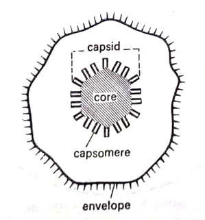
The capsid is made up of many identical structural units, the capsomeres. The composition, number, and form of capsomeres vary with the kinds of viruses. The core with its capsid is called the nucleocapsid of the virus. Many mammalian viruses have outside the capsid also an envelope or limiting membrane or mantle, usually of lipids or lipoproteins. Such viruses are also called lipoviruses. Example – Herpes virus. icosahedral and helical viruses occur in both, naked well as enveloped forms. (BSc Microbiology Viruses Notes Study Material)
The shape of the viral particles, arrangement of capsomers in the capsid, fine details of the surface structures, and the symmetry of particles are now relatively better understood due to some sophisticated techniques. For instance, electron microscopy besides giving some clue to shape provides high magnifications (several million times) of virus particles.
Another useful technique is X-ray diffraction, which is very useful for studying the arrangement of capsomeres in the capsid. Viruses in this way are studied indirectly by placing crystals of a pure virus preparation in an X-ray beam and recording the diffraction patterns produced when the X-rays are reflected from the planes of atoms in the crystal. This could reveal that the protein subunits forming the virus shell were arranged symmetrically. (BSc 2nd Year Microbiology Viruses Notes Study Material)
For example studies on TMV particles by X-ray diffraction, revealed that the capsomeres were arranged in a helix. Most small viruses which looked spherical in electron micrographs, under X-ray diffraction, however, were found to have a cubic symmetry. This suggested that they were regular polyhedrons and also members of the group of Platonic solids-solids with 4,6,8,12 and 20 sides.
Dehydrated virus particles are transparent to an electron beam, thus being invisible. Thus techniques have been developed to make the particles visible. One such technique is shadowing, where a stream of heavy-metal atoms is allowed to fall on the virus particles at an angle. The particle specimen is put in a vacuum chamber metal atoms are evaporated from a source toward the side of the chamber. With this technique, the overall shape of the virus particle may be seen, but not at all the fine details of its surface structures.
Within the past few years a simple method of staining (negative staining or negative contrast) the isolated virus particles has been very useful than shadowing in revealing details of highly magnified particles by electron microscopy. The particles are surrounded by an electron-dense material, potassium phosphotungstate. (BSc 2nd Year Microbiology Viruses Notes Study Material)
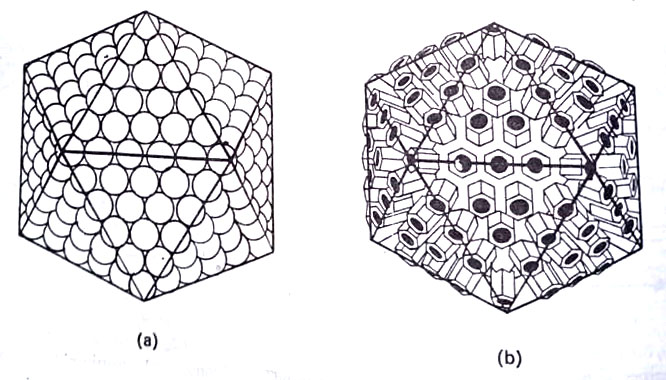
Virus suspension is mixed with a solution of potassium phosphotungstate, and this mixture is sprayed or deposited on the specimen mounts. The images produced by this method are reversed to those obtained by normal methods (hence the “negative-contrast method”). This method revealed that all viruses fall into three main symmetry groups – cubic, helical, and complex symmetry viruses.
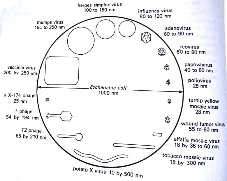
There is a considerable range in the size of viruses. Most viruses fall in a size range between 10 and 200 nm. Some like yellow fever, foot-and-mouth disease, and poliomyelitis, are small with diameters of about 25 nm, whereas others like smallpox (vaccinia) virus are large about 250 nm in diameter. Except for these large viruses, most others are invisible by optical microscope.
In outward form (shape) viruses differ widely. They may be rod-like, elongated like a piece of insulated electrical cable (TMV), rounded (mumps virus, herpes virus, influenza virus), or tadpole-like (T-even bacteriophage). The form of some viruses, especially the large pox viruses of the pox virus family as vaccinia virus are complex and often vague or indeterminable. Some viruses as rabies virus are bullet-shaped. (BSc 2nd Year Microbiology Viruses Notes Study Material)
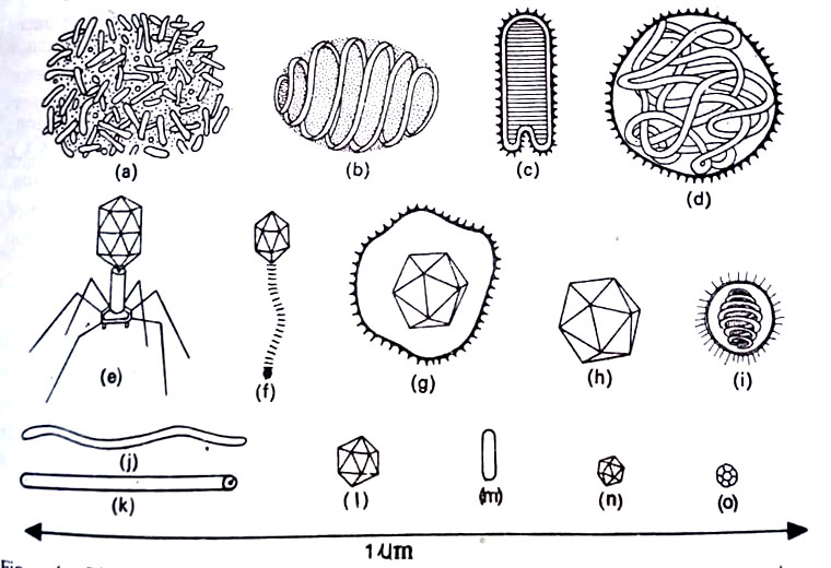
The negative staining method revealed that viruses fall into three symmetry groups. These are as follows:
1. Cubic symmetry. Examples are tipula iridescent virus (in larvae of some insects; a typical icosahedron in shape), adenovirus (associated with respiratory disease in man), herpes virus (cold sores in man), polyomavirus (tumors in rodents, cancer in a man ?), turnip yellow mosaic virus, Φ x 174 ( a bacteriophage on E. coli) and perhaps poliomyelitis virus of man.
2. Helical symmetry. Examples are the well-known plant virus tobacco mosaic virus (TMV), and some animal viruses of influenza or myxovirus group. These include viruses of mumps, new castle disease (a respiratory disease in fowl), fowl plague, and Sendai disease virus (a form of influenza).
3. Complex symmetry. Examples are the large bacterial viruses (like T2 and other T-even phages of the bacterium, E. coli) that are tadpole-shaped, and the large pox viruses of the pox virus family (viruses of variola) that include the vaccinia, cowpox, extremely and Orf viruses (contagious pustular dermatitis).
We shall consider details of the structure of some viruses belonging to each symmetry group.
Cubic Viruses
Tipula iridescent virus has the shape of a regular icosahedron. In icosahedrons, each face is an equilateral triangle. A regular icosahedron is said to have 5,3,2 symmetry. Adenovirus has a cubic symmetry. Each particle is an icosahedron. The electron micrograph shows that the surface of the particle is composed of regularly arranged structural units resembling tiny balls; the balls can be seen on the vertexes, faces, and edges of an icosahedron. (BSc 2nd Year Microbiology Viruses Notes Study Material)
There are six balls along each edge. There are 252 surface subunits or capsomeres. The herpes virus also shows a cubic symmetry exactly like an adenovirus. It has the same external shape also. However, capsomeres of the herpes virus unlike those of the adenovirus are elongated hollow prisms, some hexagonal in cross section and others pentagonal. Elongated capsomeres stand out clearly in profile at the periphery of the virus. This virus has an envelope. There are 162 capsomeres in each particle, 12 pentagonal and 150 hexagonal prisms.
The polyomavirus is smaller than the herpes virus, with fewer capsomeres. If examined by the “shadowing” technique, particles appear to be spherical. However, negative staining revealed a cubic symmetry. The shell is composed of 42 capsomeres, 12 pentagonal prisms at the corners of an icosahedron, and 30 hexagonal prisms on the 30 edges. The 20 faces have no capsomeres of their own, which helps to explain the nearly spherical appearance of the virus. (BSc 2nd Year Microbiology Viruses Notes Study Material)
Turnip yellow mosaic virus, in an electron microscope also appears to have 32 capsomeres arranged in a cubic symmetry. In X-ray diffraction pictures there appear 60 capsomeres, that possibly combine in some way to give the appearance of only 32 subunits when the particle is seen under an electron microscope. (BSc 2nd Year Microbiology Viruses Notes Study Material)
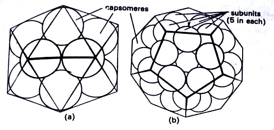
High-resolution electron micrographs revealed that structures originally identified as capsomeres are indeed composed of still smaller subunits. The virus Φ x 174 has been intensively studied because it contains an unusual single-stranded form of DNA. When first examined under an electron microscope, the virus appeared to have a shell of 12 spherical capsomeres in icosahedral symmetry. This is the minimum number needed for an icosahedron. (BSc 2nd Year Microbiology Viruses Notes Study Material)
Later, it was shown that each capsomere is formed from five subunits, but since each capsomere may be shared with a neighbor, the total number of subunits is 30. If they are not shared, and each capsomere has five subunits, arranged in a ring-like structure the total would be 60, and the shape of the particle would be that of a dodecahedron (12-faced-polyhedron).
Helical Symmetry
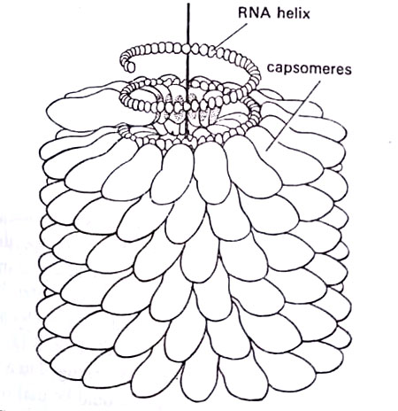
The best-known member of this group is the tobacco mosaic virus (TMV) helical structure was originally inferred from X-ray diffraction data. These data coupled with evidence from other physical and chemical studies could provide a detailed architecture of the viral particle. The capsomeres are elongated structures arranged in a helical manner. Approximately 16 capsomeres form one turn of a helix.
The subunits project from a central axial hole that runs the entire length of the virus. The nucleic acid does not occupy the hole, but is deeply embedded in the protein subunits and describes a helix of its own. The virus is composed of 2,130 identical protein subunits, each being a large molecule of 168 amino acids. (BSc 2nd Year Microbiology Viruses Notes Study Material)
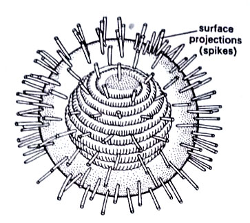
Until recently helical symmetry was observed only in plant viruses. Now it has also been seen in the complex animal viruses that are members of influenza, or myxovirus, group. The group includes the viruses of mumps new castle disease, fowl plague, and Sendai disease. Electron micrographs produced by shadow-casting showed various shapes of these viruses. Some are roughly spherical, some filamentous, and others complex and irregular.
The section of the purified virus showed an internal component in the form of a ring-like structure surrounded by an outer membrane. The negative staining method showed that the internal component or capsid had the same dimensions and appearance as the rods of TMV but is more flexible. For instance, in the mumps virus, the helical capsid forms coils or loops after being released. The particles of influenza and fowl plague are structurally more compact than the mumps virus and, unless subjected to special chemical treatments, are rarely observed releasing their internal components. (BSc 2nd Year Microbiology Viruses Notes Study Material)
The envelopes of influenza and fowl plague viruses carry functional, surface projections called spikes. The spikes contain the enzyme neuraminidase, which interacts with the cell membrane during attachment to the cell. Influenza, measles, and mumps viruses possess this enzyme. Another enzyme of the spikes, hemagglutinin, allows viruses to agglutinate, or clump, red blood cells (that is why the enzyme owes its name). Polio viruses and adenoviruses, as well as influenza, measles, and mumps viruses, have this property.
If these viruses are treated with either the internal helix is released and can be separated from hemagglutinin in a centrifuge. This inner component is of a smaller diameter than that of viruses of mumps, Newcastle disease, and Sendai disease. A possible arrangement of the components in a typical myxovirus is shown in Figure.
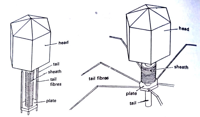
Complex Symmetry
This group includes large bacterial viruses, such as the T2 virus which infects the bacterium E. coli, and the large pox viruses. The T2 virus has a head of the shape of a bipyramidal hexagonal prism. Attached to one end of the prism is a tail structure consisting of a helical contractile sheath surrounding a central hollow core. At the extreme end of the core, there is a curious hexagonal plate carrying six slender tail fibers. The plate structure and tail fibers make initial contact with the bacterium cell wall. After contact has been made, the helical sheath contracts, allowing the nucleic acid core of the virus to enter the bacterial cell.
Still larger viruses of complex symmetry are several important members of the pox virus family – the viruses of variola, vaccinia, cowpox, and ectromelia. They may be seen with a light microscope. For instance, the vaccinia virus is about twice the diameter of the smallest living cells-the PPLOs. In an electron micrograph, the vaccinia virus appears to have a three-dimensional bricklike shape with a spherical dense central region, surrounded by a number of layers or membranes of varying opacity to the electron beam.
In some micrographs, there may be seen some tube-like structures between the outer membranes and the central region. The particles of the virus that causes Orf, or contagious pustular dermatitis, the tubular components form a definite crisscross pattern. It is difficult to say whether the tubular structures are capsids or capsomeres, and where the nucleic acid is located in relation to them.
Tobacco Mosaic Virus (TMV)
TMV is a rod-shaped virus. Each particle is elongated, cigarette-like in form. Each rod is about 3000 A (300 nm) long and of 150 (15 nm) in diameter. In each rod, there are about 2130 protein subunits – the capsomeres. The capsomeres are closely packed and arranged in a helical manner around the RNA helix: forming a hollow cylinder. (BSc 2nd Year Microbiology Viruses Notes Study Material)
Thus, there is a hollow core (axial hole) of about 4 nm (40 A) diameter which runs the entire length of the rod and it contains the RNA. The RNA does not occupy the hole but is deeply embedded in the protein subunits, thus having its own helix. RNA is a single-stranded molecule in the form of a long helix extending the entire length of the particle.
The protein amounts to about 95% of the total substances of the rod and the remaining 5% is RNA. The proteins are of high molecular weight, about 40 million, whereas the molecular weight of RNA molecules is about 2 million. RNA provides a code that directs the synthesis of specific viral proteins in the host cell. (BSc 2nd Year Microbiology Viruses Notes Study Material)
There are about 161/3 protein subunits in each helical turn, and three turns of the helix contain about 49 capsomeres. Each capsomere has a molecular weight of about 17,300 and is arranged in the virus with a pitch of 23 Å. The capsid has all amino acids found in other plant proteins. Each capsomere contains about 168 amino acid molecules. (BSc 2nd Year Microbiology Viruses Notes Study Material)
The particles were crystalized by Stanley (1935) who found that crystals were highly infectious. It was known later that RNA alone is infectious. The virion may remain infective for about 50 years and can withstand boiling for about 10 minutes.
The particles infect tobacco leaves. It is transmitted and introduced into the host cells by vectors like Myzus pseudosolani, M. circumflexes, and grass-hopper. It may also be transmitted mechanically through rubbing, transplanting, and handling. Once inside the host cell, the viral RNA directs the metabolic system of the host to synthesize its own proteins. There occurs replication of RNA molecules also inside the host cell. All the raw materials are derived from the host. Thus, there is formed a progeny of viral particles inside the host cell, are sooner or later released after lysis of the cell. (BSc 2nd Year Microbiology Viruses Notes Study Material)
TMV can infect tomatoes and many other plants also, including most of the family Solanaceae.
Geminiviruses
They multiply only in plants. They have circular single-stranded DNA of mol. wt. 0.7-1×10⁶. 18 nm quasi-isometric particles occurring in pairs are usually found in the nucleus. One molecule of DNA per pair of particles. Persistent in whitefly or leafhopper vectors.