BSc Microbiology Eukaryotic Microbes Notes Study Material
BSc Microbiology Eukaryotic Microbes Notes Study Material: BSc is a three-year program in most universities. Some of the universities also offer BSc Honours. After getting enrolled for BSc, there are certain things you require the most to get better grades/marks in BSc. Out of those, there are BSc Study Material, BSc Sample Model Practice Mock Question Answer Papers along with BSc Previous Year Papers. At gurujistudy.com you can easily get all these study materials and notes for free. Here in this post, we are happy to provide you with BSc 2nd Year Microbiology Eukaryotic Microbes Notes Study Material.

BSc Microbiology Eukaryotic Microbes Notes Study Material
Protozoa
They are a group of single-celled organisms, simple in structure, though functionally complex. More than 30,000 species have been reported which exist mostly in aquatic habitats. They are often included in zoology textbooks. They are considered in microbiology due to their unicellular structure, microscopic size, and as agents of human diseases. The discipline concerned with medically related protozoa and multicellular parasites is called parasitology. (BSc Microbiology Eukaryotic Microbes Notes Study Material)
Characteristics of Protozoa
(1) Largest organisms included in the microbial world.
(2) Except for a few, they lack chlorophyll or other photosynthetic pigments.
(3) Though unicellular, but able to perform all functions characteristic of multicellular organisms.
(4) Mostly live in water, damp soil or mud, in drainage ditches or puddles, in ponds or oceans.
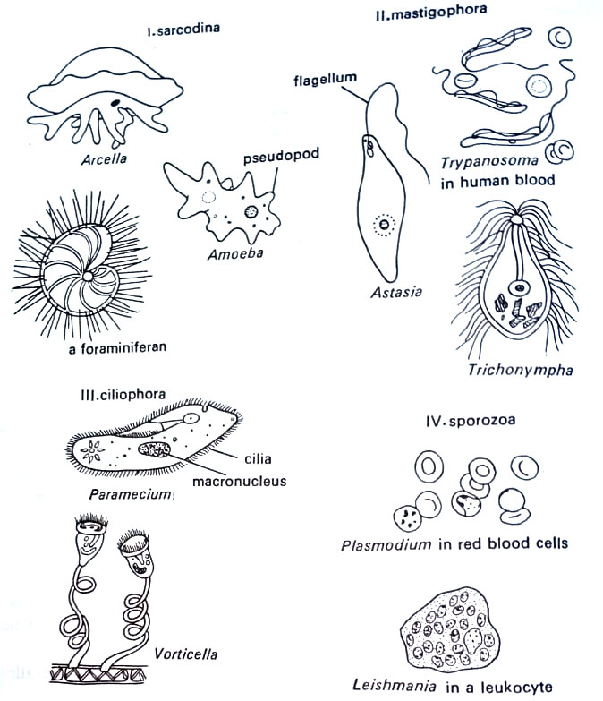
(5) A great diversity in form as shown in Figure.
(6) Cells enclosed by a membrane which in some cases surrounded by a pellicle containing chitinlike material for rigidity. The cell wall, however, is absent. True nuclei, flagella, and cilia are present. Freshwater forms take in water by osmosis and eliminate it via organelles called contractile vacuoles.
(7) Nutrition by ingestion i.e. engulfing food particles by phagocytosis or through special organs of ingestion. A membrane encloses the particle, forming a food vacuole. The latter commonly joins with a submicroscopic organelle, lysosome which contains digestive enzymes.
(8) Generally heterotrophic, saprobic, and pathogenic. The feeding form is commonly known as the trophozoite.
(9) Aerobic.
In Whittaker’s system, they along with certain algae are placed in the kingdom Protista. Four major classes are recognized.
(1) class Sarcodina (Amoebae)
(2) class Mastigophora (Flagellates)
(3) class Ciliophora (Ciliates)
(4) class Sporozoa (Nonmotile forms)
Sarcodina. This class includes amoebae, in which movement is by pseudopodia. These are projections of the cell membrane into which the cytoplasm flows slowly. The motion is called amoeboid motion. Reproduction is by binary fission preceded by a duplication of the nucleus. The feeding pattern is phagocytosis. (BSc Microbiology Eukaryotic Microbes Notes Study Material)
Two large groups of marine amoebas are included in this class (i) Radiolaria, which are abundant in the Indian and Pacific oceans; have spherical shells. (ii) The second group called Foraminifera has snail-like shells and live in hard shell-like coverings composed of calcium carbonate. Amoebae also serve as research tools due to their large size and ease of cultivation. Their nucleus can be easily removed by microsurgery and transplanted into other cells. They cause amoebiasis and other infections.
Mastigophora. They possess one or more whip-like, undulating flagella to propel the cell forward. The structure of the flagellum is similar to that of a sperm cell flagellum, with a typical 9+2 arrangement. Undulations sweep the flagella to the tip, and the lashing motion forces water outward to provide locomotion. This class includes the largest (about 50%) collection of protozoa. A common example is a green flagellate, Euglena gracilis, shown in Figure.
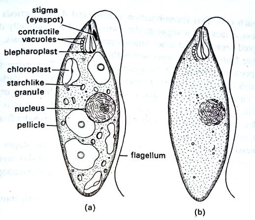
It has a single nucleus and flagellum, and an elongated cell structure. It occurs in freshwater ponds. It contains chloroplasts in the cytoplasm, thus photosynthetic. Another example of a similar type is Astasia longa. This group also includes some important human parasites infecting the nervous, urogenital, and intestinal systems. (BSc Microbiology Eukaryotic Microbes Notes Study Material)
Ciliophora. They have complex cells that range in size from 10 µm to 3 mm. They are characterized by rows of hair-like appendages, the cilia, which structurally resemble flagella showing a 9+2 arrangement. However, they do not lash about. Rather, they beat synchronously similar to the motion of the teeth of a comb as you pass your finger down the row. This provides a smooth, even motion in contrast to the jerky motion of the flagellates.
Paramecium is slipper-shaped with a primitive gullet and mouth into which food particles are swept by cilia. There is a single large macronucleus to one or more micronuclei. During sexual conjugation, two cells make contact, and a cytoplasmic bridge forms between them. A micronucleus in each cell undergoes two divisions to form four micronuclei per cell.
Three micronuclei disintegrate and the remaining one undergoes division. An exchange of micronuclei takes place. The cells separate, the micronuclei fuse, and a new micronucleus forms in each cell. This recombination is observed during periods of environmental stress, a factor that suggests the formation of a genetically different and better-adapted organism. At other times reproduction is by mitosis.
Another feature of Paramecium is the kappa factor. These nucleic acid particles appear responsible for the synthesis of toxins. The toxins destroy those ciliates which lack these factors. Evidence suggests that these particles may be bacteria or viruses. Paramecium species also possess trichocysts – organelles that discharge filaments and trap the organism’s prey. Another feature is the contractile vacuoles, the bubble-like organelles which pump excess water from the cytoplasm to the external environment. Some are pathogenic.
Sporozoa. Their life cycles are very complex. Some stages in the life cycle resemble bacterial or fungal spores due to which they owe this name. They are non-motile; occasionally, forms in the life cycle have flagella or pseudopodia, which are short-lived. Adults are non-motile.
Most members of this group are parasites. Two members, those of malaria and toxoplasmosis are noteworthy.
Amoebiasis. The disease is characterized by painful ulcers of the human intestine. The causal organism is Entamoeba histolytica (Sarcodina). It enters the body as a cyst and passes through the stomach acid, as shown in Figure.
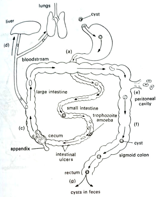
In small and large intestines the amoebae emerge and begin to feed on tissue. This feeding form is known as the trophozoite. Lesions coalesce to form a deep ulcer, causing appendicitis-like pain. If not controlled, the amoebae eventually reach the blood vessels of the intestinal wall. Now the stool becomes bloody, causing the problem of parasitemia. If amoebae localize in the lung or liver, these may be fatal. Amoebiasis may be treated with paromomycin and metronidazole and tetracycline.
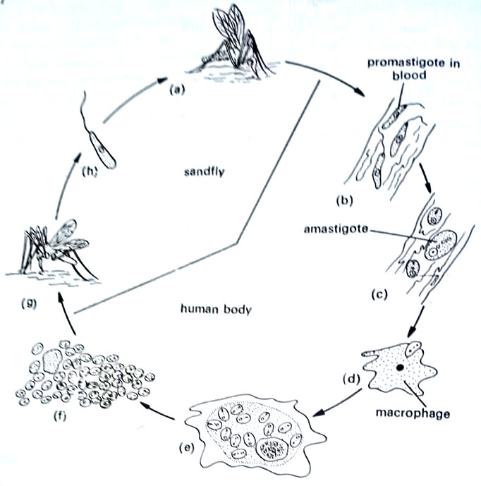
Leishmaniasis (kala-azar). The causal organism is Leishmania donovani (Mastigophora). It occurs in two stages: the non-flagellated amastigote or Leishman-Donovan body, and the flagellated promastigote or cryptomonad stage. The first occurs in humans, and the second in the insect vector.
Kala-azar (black fever) is transmitted by the sandfly of the genus Phlebotomus which initially acquires the protozoa when it takes a blood meal from an infected patient. The parasites fill the insect’s gut and then pass to the salivary Eland from which they are injected into the next victim.
The disease is characterized by infection of the phagocytes of the body’s reticuloendothelial system (RES). These include the white blood cells and various tissue phagocytes called macrophages, which are found in the spleen, liver and lymph nodes, and other organs. When the phagocytes engulf the parasites, the latter multiply within the phagocyte’s cytoplasm causing it to burst. The new parasites are then engulfed by other phagocytes and the cycle is repeated. (BSc Microbiology Eukaryotic Microbes Notes Study Material)
Symptoms are fever, swelling of lymph nodes, especially in the neck, and progressive anemia, weakness, and emaciation. As the number of phagocytes declines the bone marrow attempts to replace them at the expense of other white blood cells. The lowered white blood cell count is referred to as leukopenia.
The treatment includes antimony compounds and either of two normally antifungal agents, nystatin or amphotericin B.
Malaria. The disease was described as early as the 5th century B.C. There are four recognized species of Plasmodium that are related to malaria: P. vivax, ovale. P. malariae and P. falciparum, all transmitted by female Anopheles mosquitoes. This insect consumes human blood to acquire a component for the production of her eggs. (BSc Microbiology Eukaryotic Microbes Notes Study Material)
The parasite has a very complex life cycle. It takes place partly in mosquitoes and partly in human blood. The parasite enters the bloodstream in the sporozoite form and immediately invades the liver, which is very difficult to treat.
After several hours, many of the merozoites emerge and penetrate the red blood cells. Following a complex series of transformations, thousands of RBCs rupture simultaneously and release tens of thousands of new parasites. At this point, the victim suffers a malaria attack. There is severe cold, the temperature rises rapidly to 104°F – 106°F and there is a severe headache with mild delirium. After the next two or three hours, massive perspiration ends the hot stage. The patient has a sound sleep, till the next attack.
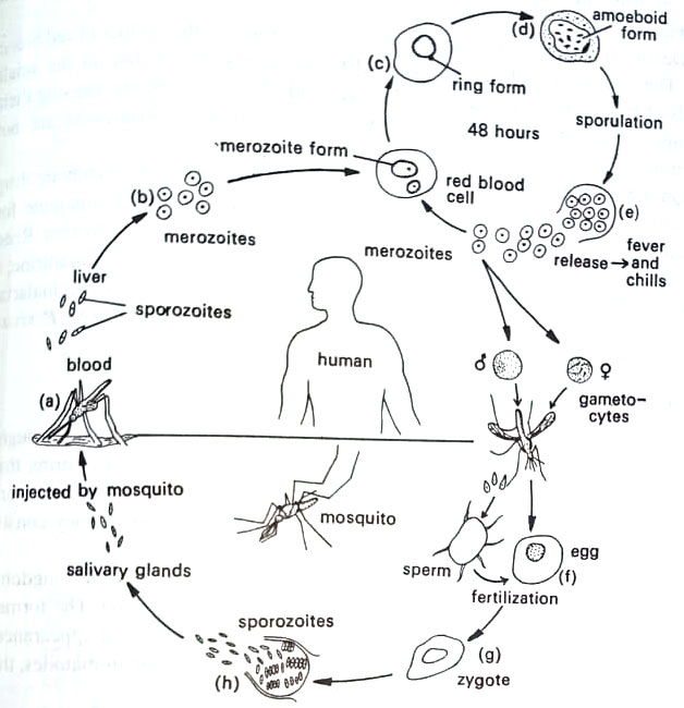
During this period, the parasite enters a new set of RBCs and repeats the transformation cycle. Plasmodium vivax and P. ovale spend about 48 hr in RBCs and there is a 48 hr interval between attacks. This malaria is called tertian malaria (three days). For P. malaria, the cycle is about 72 hr – therefore, quartan malaria (four days). The cycle of P. falciparum is not defined and attack may occur at irregular intervals. This type of malaria is called estivo-autumnal malaria.
Death from malaria may be due to several factors, related to the loss of red blood cells. The anemia is substantial. The RBC fragments accumulate in the small vessels of the brain, kidneys, heart muscle, liver, and other vital organs, causing their blockage. Heart attacks, cerebral hemorrhages, and venal shutdowns are not uncommon. (BSc Microbiology Eukaryotic Microbes Notes Study Material)
Quinine is used to treat malaria since its discovery in 1640. Synthetic drug chloroquine is also used for the clinical phase of malaria, and primaquine for the dormant phase. Recently mefloquine is also used. Most recently Walter Reed Army Institute for Research in Washington, U.S.A. has developed halofantrine, a new anti-malaria drug that is effective in all forms of multidrug-resistant malaria Halfan, as the compound is known, is shown to cure more than 95% of both, P. vivax and P. falciparum.
Multicellular Parasites of Humans
These parasites are the roundworms and flatworms, commonly found throughout the world. They range in size from tiny flukes to tapeworms that sometimes reach 20 feet in length. Due to their infectious nature, they are considered in microbiology. Together with the unicellular protozoa, they constitute the discipline known as parasitology.
Multicellular parasites are grouped into two phyla of the animal kingdoms: Platyhelminthes (flatworms) and nemathelminthes (roundworms). The former has a broad or flat body, whereas the latter has a long threadlike appearance. The flatworms include the cestodes, known as tapeworms, and the trematodes, the flukes. (BSc Microbiology Eukaryotic Microbes Notes Study Material)
The life cycle of parasitic worms involves several hosts. The final host – the definitive host is the one in which adult sexually mature is developed. We shall consider here mainly those worms whose definitive host is human. The adults usually produce eggs that are released from the body after fertilization. The eggs develop into larvae in one or more intermediate hosts. The cycle is completed when the larvae reach again to the human body by water or soil contact or by consumption of contaminated food or water.
Roundworms
The roundworms or nematodes vary from the tiny Trichinella worm, the cause of trichinosis, to the foot-long Ascaris, like an earthworm. Some are threadlike as filarial worms, and others such as hookworm are more rounded. Most of these live in human intestines and reproduce sexually by forming eggs, which are often excreted into the environment. After the development of larvae, the parasite is passed to the next victim. Filarial worm depends on mosquito for transfer.
Pinworm disease. This is caused by a common nematode, Enterobius vermicularis, and the most common parasite of humans. The female is about 10 mm long and the male of about half that size. Both live in the latter part of the small intestine and in the large intestine. Typical symptoms are diarrhea.
The parasite releases thousands of fertilized eggs by females to the region of the anus. Scratching the area brings the eggs to the nails and fingertips causing reinfection through the mouth. Others may be infected from eggs deposited in food, or on clothing or bed linens. Usually, the anal area itches intensely, which is a common sign of disease.
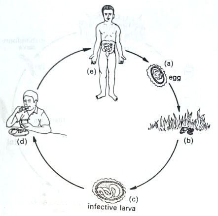
Roundworm disease. This is commonly caused by Ascaris lumbricoides, the largest of intestinal nematodes. Tropical and subtropical areas are the common foci of this disease. The nematode is like an earthworm. The female is over a foot long and the male is somewhat smaller with a curled tail. Heavy infection may cause intestinal obstruction due to the presence of tightly compacted masses of the parasite.
The female is a prolific egg producer, sometimes producing 200,000 per day. After fertilization, the eggs are excreted into the soil where they hatch into larvae. These attach to plants from which are again ingested. In many parts of the world human feces – night soil is used as a fertilizer for vegetables. This helps in the spread of the parasite. Water pollution from the soil and contact with contaminated fingers are other ways of transmission. Roundworm larvae may also pass from the intestine to other parts of the body. They may be passed to the lungs causing a pneumonialike disorder. (BSc Microbiology Eukaryotic Microbes Notes Study Material)
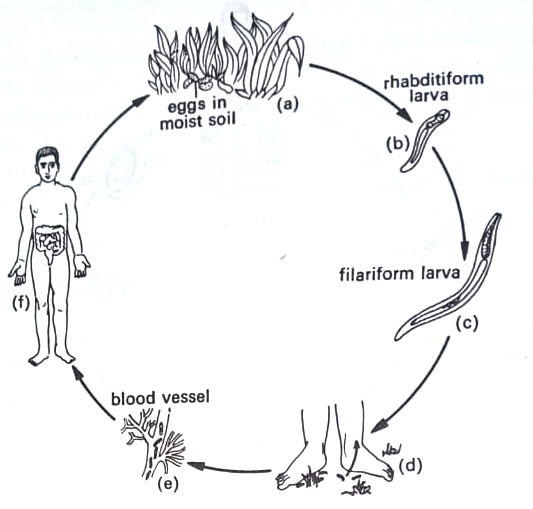
Hookworm disease. This is caused by two nematodes: the old world hookworm, Ancylostoma duodenale, and the New World hookworm, Necator americanus. Of these, the former is common in Europe, Asia, and the U.S.A. Both are about 10 mm long with a set of hooks or sucker devices, that allow them to attach firmly to the intestine. They may induce ulcers and consume blood, causing anemia.
Hookworm eggs pass into the soil where the larvae emerge as long rhabditi-form larvae. These convert to hair-like filariform larvae. The filariform larvae attach to the skin during contact with moist vegetation in the soil and penetrate the skin layers to the bloodstream. They localize in the lungs and are carried up the bronchi, from where they are swallowed. The infection then occurs in the intestine.
Filiariasis. The worm of this disease is a threadlike nematode – Wuchereria bancrofti, present in hot, humid climates. The worm breeds in tissues of the circulatory system causing extensive inflammation and damage to lymphatic vessels. If the parasite persists longer in the body, the legs or scrotum may become swollen and distorted with lymph fluid. A condition called elephantiasis may develop.
The females are about 100 mm long and the males are about half that length. Within the female, fertilized eggs give tiny eel-like embryos called microfilariae. These enter the bloodstream and are ingested by mosquitos during a blood meal. They undergo further development in the muscle of the mosquito and are injected back into humans during blood meals. The adults are formed in lymph vessels to establish infection. (BSc Microbiology Eukaryotic Microbes Notes Study Material)
Flatworms
They belong to the phylum Platyhelminthes. The intestinal tract of these worms consists of a blind sac. The single worm contains both male and female sex organs (hermaphroditic). The life cycle is rather complex involving several different forms and a number of intermediate hosts. Human intermediate hosts.
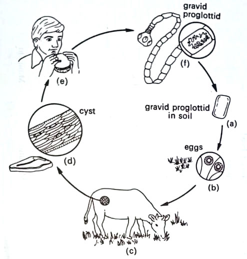
The tapeworms (cestodes) consist of a head region called a scolex, a neck, and a series of segments called proglottids. The scolex holds tightly to the host tissue, and new proglottids are produced in the neck area. Sex organs are located in mature proglottids in the center of the worm. The most distal proglottids, called gravid proglottids, are filled with fertilized eggs.
The flukes (trematodes) are broad leaf-like with oral suckers for attachment to host tissue. From fertilized eggs, many ciliated forms called miracidium to develop. The miracidium penetrates a snail where it is transformed into a cercaria. It is then released as an encysted metacercaria and makes its way back to the human in this form. (BSc Microbiology Eukaryotic Microbes Notes Study Material)
Beef and pork tapeworm diseases. Human is the definitive host for both the beef tapeworm, Taenia saginata, and the pork tapeworm T. solium. The former is about 25 feet long, whereas the latter is about 8 feet long with up to 2,000 proglottids. The main damage is caused to the intestine, causing an obstruction. The life cycle involves the release of gravid proglottids to the soil, from where they are consumed by cattle or pigs. Embryos are released from the eggs and these are passed to the muscle of animals where they encyst. These may then be consumed in poorly cooked beef or pork. (BSc Microbiology Eukaryotic Microbes Notes Study Material)
Liver fluke disease. This is caused by Fasciola hepatica, a leaf-like flat worm, commonly found in sheep and cattle. Eggs from the animal may be deposited in water. If snails are present, the conversion from miracidium to cercaria to metacercaria may occur.
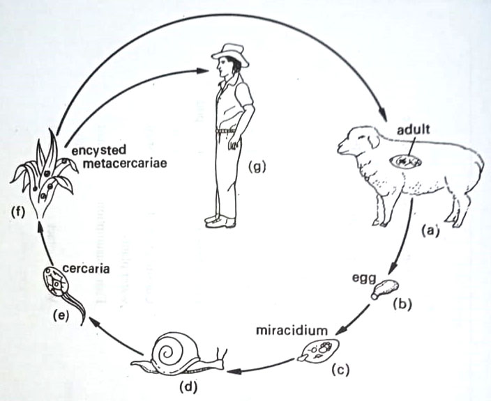
The cysts collected on vegetation may be then ingested by humans. The intestinal wall is penetrated, followed by the migration of the parasite to the liver where it localizes. There may be significant damage to the liver. Liver flukes are common in Far East Countries.
BSc Microbiology Eukaryotic Microbes Notes Study Material
BSc 2nd Year Sample Model Practice Mock Test Question Answer Papers